Request Demo
Best Practices for Lentiviral Vector Concentration and Titration
9 May 2025
Lentiviral vectors are widely used tools in molecular biology for the delivery of genetic material into cells. Their ability to integrate into the host genome makes them particularly valuable for stable gene expression. However, to achieve optimal results, it is crucial to concentrate and titrate these vectors properly. Below are best practices that can guide researchers through these processes effectively.
Firstly, it is essential to start with a high-quality lentiviral vector preparation. The vector should be produced using a well-optimized protocol that minimizes contaminants and maximizes viral particle yield. This typically involves transfecting a suitable producer cell line, such as HEK293T cells, with a combination of plasmids encoding the viral components and the gene of interest.
Once the lentiviral particles are produced, concentration is often necessary to achieve the titers required for efficient transduction. One of the most common methods for concentrating lentiviral vectors is ultracentrifugation. This method involves centrifuging the viral supernatant at high speeds, allowing the viral particles to form a pellet at the bottom of the tube. The supernatant is carefully removed, and the pellet is resuspended in a smaller volume to achieve the desired concentration. It is important to perform this step under sterile conditions to prevent contamination.
An alternative to ultracentrifugation is the use of polyethylene glycol (PEG) precipitation. This method involves adding PEG to the viral supernatant, which causes the viral particles to precipitate out of solution. Following incubation and low-speed centrifugation, the precipitated viral particles can be resuspended in a smaller volume. PEG precipitation is generally less labor-intensive than ultracentrifugation and can be scaled up more easily.
After concentrating the lentiviral vectors, accurate titration is crucial to determine the functional titer of the viral preparation. One widely used method is the quantitative polymerase chain reaction (qPCR) to measure the number of viral genomes. This method provides a quick and relatively accurate estimation of the viral titer. However, it is important to note that qPCR does not differentiate between functional and non-functional viral particles.
Another method for titration is the functional assay, where the concentrated virus is used to transduce a permissive cell line, such as HEK293T or HeLa cells. After transduction, the expression of the reporter gene or marker is measured, typically by flow cytometry or fluorescence microscopy, to determine the number of transducing units per milliliter. This method provides a direct measure of the functional titer and is considered more representative of the virus’s transducing capacity.
It is also recommended to include a negative control in the titration assays to account for any background signal. Additionally, performing titration in multiple replicates can help ensure the accuracy and reliability of the results.
Finally, it is crucial to store the concentrated lentiviral vectors appropriately to maintain their stability and functionality. Aliquot the concentrated virus to avoid repeated freeze-thaw cycles, which can significantly reduce viral titer. Store the aliquots at ultra-low temperatures, typically -80°C, for long-term storage.
In summary, the concentration and titration of lentiviral vectors are critical steps in the successful application of these tools in research and therapeutic settings. By following best practices, including high-quality vector preparation, using suitable concentration methods, and performing accurate titration assays, researchers can maximize the efficacy and reliability of their lentiviral-based experiments.
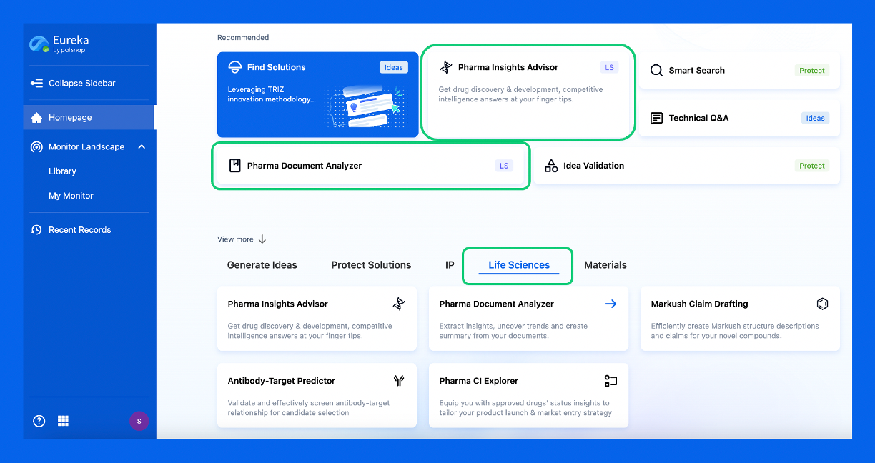
Discover Eureka LS: AI Agents Built for Biopharma Efficiency
Stop wasting time on biopharma busywork. Meet Eureka LS - your AI agent squad for drug discovery.
▶ See how 50+ research teams saved 300+ hours/month
From reducing screening time to simplifying Markush drafting, our AI Agents are ready to deliver immediate value. Explore Eureka LS today and unlock powerful capabilities that help you innovate with confidence.

AI Agents Built for Biopharma Breakthroughs
Accelerate discovery. Empower decisions. Transform outcomes.
Get started for free today!
Accelerate Strategic R&D decision making with Synapse, PatSnap’s AI-powered Connected Innovation Intelligence Platform Built for Life Sciences Professionals.
Start your data trial now!
Synapse data is also accessible to external entities via APIs or data packages. Empower better decisions with the latest in pharmaceutical intelligence.