Request Demo
Compare ECL vs. Fluorescent Detection in Western Blotting
9 May 2025
Western blotting is a widely used analytical technique in molecular biology and biochemistry, employed to detect specific proteins in a sample. Two popular methods for visualizing proteins in western blotting are Enhanced Chemiluminescence (ECL) and fluorescent detection. Both techniques have their own advantages and limitations, making the choice between them dependent on the specific needs of the experiment. In this blog, we will compare ECL and fluorescent detection, exploring their principles, benefits, and drawbacks to help you make an informed decision for your laboratory work.
Enhanced Chemiluminescence (ECL) is a traditional and highly sensitive method for detecting proteins on a western blot. It relies on the emission of light during a chemical reaction. Typically, after the protein transfer and blocking steps, the membrane is probed with a primary antibody specific to the target protein. A secondary antibody conjugated to an enzyme such as horseradish peroxidase (HRP) is then applied. When the ECL substrate is added, HRP catalyzes a reaction that produces light, which can be captured on film or by a digital imaging system.
One of the key advantages of ECL is its high sensitivity, capable of detecting low-abundance proteins. It is also relatively cost-effective and does not require specialized equipment beyond a darkroom or imaging system. However, the signal generated by ECL is transient, typically lasting only a few hours, which limits the opportunity for repeated analysis. Additionally, the dynamic range of ECL detection can be limited, making it challenging to quantify proteins accurately, especially across samples with varying expression levels.
In contrast, fluorescent detection in western blotting uses antibodies conjugated to fluorescent dyes. After the transfer and blocking steps, the membrane is incubated with a primary antibody and then with a fluorescently labeled secondary antibody. The bound antibodies can be visualized and quantified using a fluorescence scanner or imager.
Fluorescent detection offers several advantages over ECL. It provides a broad dynamic range and allows for precise quantification of protein levels. The fluorescent signal is stable over time, enabling the membrane to be re-scanned without significant signal loss, thus facilitating repeated measurements and long-term data storage. Moreover, it permits multiplexing, allowing for the simultaneous detection of multiple proteins in a single blot if different fluorophores are used.
However, fluorescent detection also has its drawbacks. The initial cost of acquiring a fluorescence imaging system can be high, and the fluorescently labeled antibodies are generally more expensive than their HRP-conjugated counterparts. Additionally, fluorescent detection can suffer from background fluorescence or bleed-through between channels if multiplexing is not carefully controlled.
When considering which method to use, several factors should be taken into account. If sensitivity and cost-effectiveness are your primary concerns, ECL might be the preferred choice. On the other hand, if you require quantitative data, multiplexing capabilities, and stable signals for long-term analysis, fluorescent detection may be more suitable.
In conclusion, both ECL and fluorescent detection have their distinct advantages and limitations in western blotting applications. The decision between them depends on the specific requirements of your experiment, including sensitivity, cost, quantification needs, and the availability of equipment. Understanding these differences will enable you to select the method that best aligns with your research goals, ensuring reliable and reproducible results in your protein studies.
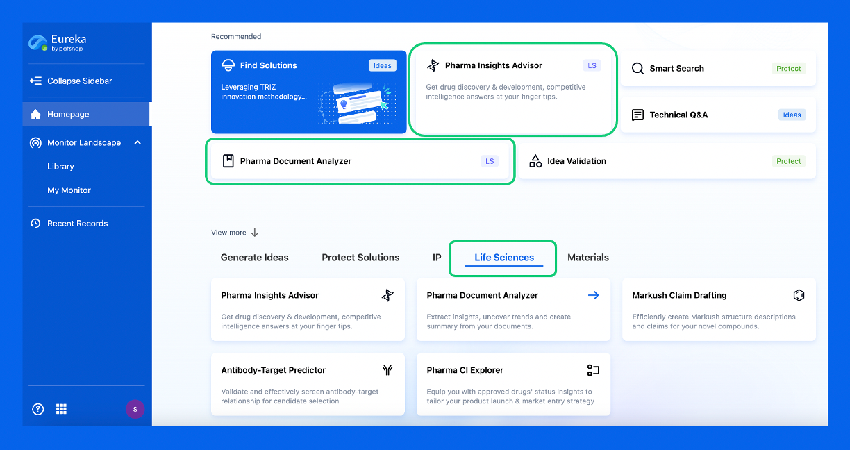
Discover Eureka LS: AI Agents Built for Biopharma Efficiency
Stop wasting time on biopharma busywork. Meet Eureka LS - your AI agent squad for drug discovery.
▶ See how 50+ research teams saved 300+ hours/month
From reducing screening time to simplifying Markush drafting, our AI Agents are ready to deliver immediate value. Explore Eureka LS today and unlock powerful capabilities that help you innovate with confidence.

AI Agents Built for Biopharma Breakthroughs
Accelerate discovery. Empower decisions. Transform outcomes.
Get started for free today!
Accelerate Strategic R&D decision making with Synapse, PatSnap’s AI-powered Connected Innovation Intelligence Platform Built for Life Sciences Professionals.
Start your data trial now!
Synapse data is also accessible to external entities via APIs or data packages. Empower better decisions with the latest in pharmaceutical intelligence.