Request Demo
EEG vs fMRI: Which Brain Imaging Method Should You Use?
28 May 2025
Understanding Brain Imaging Techniques
When it comes to exploring the brain's intricate functions and understanding neurological processes, brain imaging techniques play a crucial role. Two of the most commonly used methods are Electroencephalography (EEG) and Functional Magnetic Resonance Imaging (fMRI). Each has its unique advantages and limitations, which often makes choosing the right method a complex decision. This blog will delve into the strengths and weaknesses of EEG and fMRI, helping you make an informed choice for your research or clinical needs.
EEG - Unveiling Electrical Activity
EEG is a non-invasive technique that records electrical activity in the brain using electrodes placed on the scalp. This method is renowned for its excellent temporal resolution, capturing rapid changes in brain activity in real-time. EEG is particularly useful in situations where timing is crucial, such as diagnosing epilepsy, sleep disorders, and studying cognitive processes during specific tasks.
Advantages:
1. **Temporal Resolution**: EEG can detect changes in brain activity within milliseconds, making it ideal for studies requiring precise timing.
2. **Cost-effective**: Compared to fMRI, EEG equipment is generally less expensive and more accessible, which can be beneficial for large-scale studies or when budget constraints are a concern.
3. **Portability**: EEG devices are relatively portable and can be used in various settings, from clinical environments to research labs.
Limitations:
1. **Spatial Resolution**: EEG has limited ability to pinpoint the exact location of brain activity, which can be a drawback when spatial precision is necessary.
2. **Sensitivity to Artifacts**: EEG recordings can be influenced by external factors such as muscle movement or electrical interference, potentially complicating data interpretation.
fMRI - Mapping Brain Activity
fMRI measures brain activity by detecting changes in blood flow, offering insights into the brain's functional architecture. This imaging technique boasts impressive spatial resolution, allowing researchers to localize brain activity with precision. fMRI is particularly valuable for understanding how different brain regions contribute to various cognitive and emotional processes.
Advantages:
1. **Spatial Resolution**: fMRI provides detailed images of brain structures, allowing for accurate localization of activity.
2. **Comprehensive Data**: This method can offer a wider view of the brain’s overall functionality, making it suitable for complex cognitive and behavioral studies.
3. **Non-invasive**: Like EEG, fMRI is non-invasive, making it safe for repeated use in both clinical and research settings.
Limitations:
1. **Temporal Resolution**: fMRI cannot match the rapid temporal precision of EEG, as it detects changes over several seconds.
2. **Cost**: The equipment and operation costs for fMRI are significantly higher than those for EEG, often limiting its availability.
3. **Restrictions**: Participants must remain completely still during scanning, which can be challenging for some populations, including children or individuals with movement disorders.
Choosing Between EEG and fMRI
The decision between EEG and fMRI largely depends on your research goals and constraints. If your primary focus is understanding the timing of brain processes, EEG’s superior temporal resolution may be the best choice. On the other hand, if spatial accuracy and detailed imaging are paramount, fMRI could offer the insights you need.
Combining EEG and fMRI
Increasingly, researchers are combining EEG and fMRI to leverage the strengths of both methods. By integrating EEG’s real-time monitoring with fMRI’s spatial precision, one can gain a comprehensive view of brain activity. This hybrid approach can be particularly beneficial in complex experimental designs, offering a more complete understanding of neurophysiological processes.
Conclusion
Both EEG and fMRI have their distinct advantages and are invaluable tools in the field of brain imaging. Your choice should be guided by the specific requirements of your study and the questions you aim to answer. Whether you opt for the temporal detail of EEG or the spatial clarity of fMRI, understanding the capabilities and limitations of each method will ultimately enhance the quality and impact of your research.
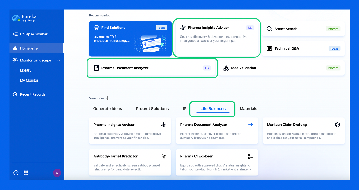
Discover Eureka LS: AI Agents Built for Biopharma Efficiency
Stop wasting time on biopharma busywork. Meet Eureka LS - your AI agent squad for drug discovery.
▶ See how 50+ research teams saved 300+ hours/month
From reducing screening time to simplifying Markush drafting, our AI Agents are ready to deliver immediate value. Explore Eureka LS today and unlock powerful capabilities that help you innovate with confidence.

AI Agents Built for Biopharma Breakthroughs
Accelerate discovery. Empower decisions. Transform outcomes.
Get started for free today!
Accelerate Strategic R&D decision making with Synapse, PatSnap’s AI-powered Connected Innovation Intelligence Platform Built for Life Sciences Professionals.
Start your data trial now!
Synapse data is also accessible to external entities via APIs or data packages. Empower better decisions with the latest in pharmaceutical intelligence.