Request Demo
How do you isolate PBMCs from blood?
28 May 2025
Introduction
Peripheral Blood Mononuclear Cells (PBMCs) are crucial components in many fields of biomedical research, including immunology, infectious diseases, and hematological studies. Isolating PBMCs from whole blood is a common laboratory procedure that allows researchers to study these cells in detail. This blog will guide you through the process of isolating PBMCs using density gradient centrifugation, a widely used method that ensures high purity and yield.
Materials Required
To begin the isolation process, you will need the following materials:
- Fresh human blood or anticoagulated blood sample (commonly with EDTA or heparin)
- Ficoll-Paque or a similar density gradient medium
- Centrifuge tubes (typically 15 ml or 50 ml)
- Sterile pipettes and pipette tips
- Phosphate-buffered saline (PBS) or another suitable washing buffer
- Centrifuge capable of maintaining a temperature of 20-25°C
- Sterile cell culture hood and appropriate personal protective equipment (PPE)
Step-by-step Procedure
1. **Sample Preparation**
Begin by gently mixing the blood sample to ensure uniform distribution of cells and anticoagulants. This can be achieved by inverting the collection tube several times. If the sample has been stored in a cool environment, allow it to reach room temperature before proceeding.
2. **Layering the Blood**
Carefully layer the blood over the Ficoll-Paque solution in a centrifuge tube. This is achieved by slowly pipetting the blood down the side of the tube to avoid mixing. The goal is to create a distinct interface between the blood and the Ficoll-Paque. The typical ratio is 2:1 of blood to Ficoll-Paque.
3. **Centrifugation**
Place the tubes in a centrifuge and centrifuge them at 400-500 x g for 30-40 minutes at room temperature. Ensure the centrifuge is properly balanced. The low acceleration and deceleration settings prevent mixing of the layers. During this step, the PBMCs will move to the interface between the plasma and the Ficoll-Paque due to their density.
4. **Harvesting PBMCs**
After centrifugation, you will observe several distinct layers. From top to bottom: plasma, a white cloudy layer containing PBMCs, Ficoll-Paque, and red blood cells with granulocytes. Carefully collect the PBMC layer using a pipette without disturbing the other layers.
5. **Washing the Cells**
Transfer the harvested PBMCs to a new centrifuge tube and dilute them with an equal volume of PBS or another suitable buffer. Centrifuge at 300 x g for 10 minutes to pellet the cells. Carefully remove the supernatant and repeat the wash step at least once more to remove any residual Ficoll-Paque and plasma.
6. **Cell Count and Viability**
After the final wash, resuspend the PBMC pellet in an appropriate volume of buffer. Count the cells using a hemocytometer or an automated cell counter. Assess cell viability using trypan blue exclusion or a similar viability dye.
7. **Storage and Application**
Depending on your experimental needs, PBMCs can be immediately used for downstream applications or cryopreserved for future use. If cryopreserving, resuspend the cells in a freezing medium (usually containing DMSO and serum) and store them in a controlled-rate freezer before transferring to liquid nitrogen storage.
Troubleshooting Tips
- Ensure the blood sample is fresh for optimal PBMC yield and viability.
- Avoid vigorous pipetting to minimize cell damage.
- If the PBMC layer is difficult to distinguish, verify that the density gradient medium and centrifuge settings are appropriate.
Conclusion
Isolating PBMCs from blood is a fundamental technique in many research labs. By following these steps carefully, researchers can obtain high-quality PBMCs suitable for a wide range of applications. Understanding the nuances of each step and taking precautions to minimize cell damage are key to successful isolation and downstream experimental success.
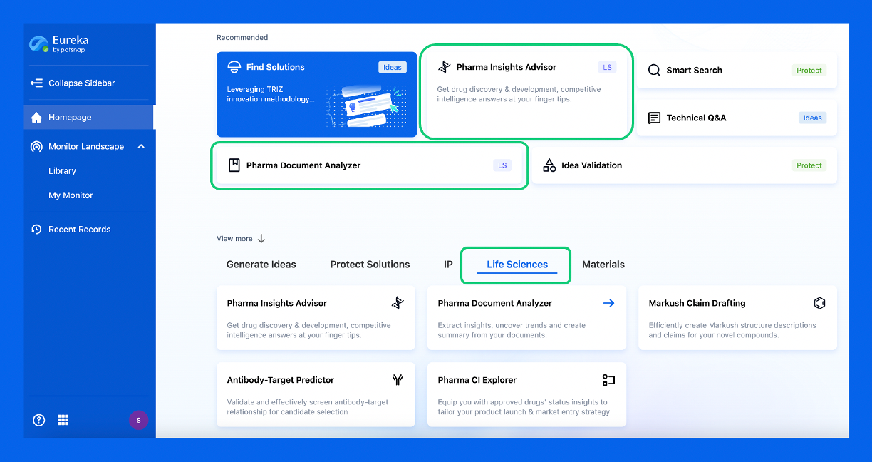
Discover Eureka LS: AI Agents Built for Biopharma Efficiency
Stop wasting time on biopharma busywork. Meet Eureka LS - your AI agent squad for drug discovery.
▶ See how 50+ research teams saved 300+ hours/month
From reducing screening time to simplifying Markush drafting, our AI Agents are ready to deliver immediate value. Explore Eureka LS today and unlock powerful capabilities that help you innovate with confidence.

AI Agents Built for Biopharma Breakthroughs
Accelerate discovery. Empower decisions. Transform outcomes.
Get started for free today!
Accelerate Strategic R&D decision making with Synapse, PatSnap’s AI-powered Connected Innovation Intelligence Platform Built for Life Sciences Professionals.
Start your data trial now!
Synapse data is also accessible to external entities via APIs or data packages. Empower better decisions with the latest in pharmaceutical intelligence.