Request Demo
How to Block a Membrane to Reduce Non-Specific Binding
9 May 2025
Blocking a membrane to reduce non-specific binding is a crucial step in various laboratory protocols, such as Western blotting, ELISA, and immunohistochemistry. Non-specific binding can lead to high background noise, which hampers the accurate detection of target proteins or antibodies. To achieve reliable results, it is essential to employ effective blocking techniques. This article will guide you through the key steps and considerations to effectively block a membrane and minimize non-specific binding.
First and foremost, understanding the nature of the membrane being used is vital. Commonly used membranes include nitrocellulose and polyvinylidene difluoride (PVDF). Each type of membrane has unique properties that can affect the blocking process. Nitrocellulose membranes are known for their high protein binding capacity, making them suitable for a wide range of applications. On the other hand, PVDF membranes are chemically resistant and have a higher binding affinity, which can sometimes lead to increased non-specific interactions. Choosing the right membrane based on your specific application and target molecules is the first step in reducing non-specific binding.
The choice of blocking agent is another critical factor. Blocking agents are typically proteins or detergents that cover the membrane surface, preventing non-specific interactions. Commonly used blocking agents include bovine serum albumin (BSA), non-fat dry milk, and casein. BSA is a highly purified protein that provides a consistent blocking effect, while non-fat dry milk is cost-effective and widely available. Casein, derived from milk, is particularly useful when working with phosphoproteins, as it contains phosphoproteins naturally. Selecting the appropriate blocking agent depends on factors such as the nature of the target protein, the detection method, and the specific requirements of the experiment.
Once the blocking agent is selected, it is essential to optimize the blocking conditions. This involves determining the appropriate concentration and incubation time. Typically, a 3-5% solution of the blocking agent is prepared in a suitable buffer, such as Tris-buffered saline (TBS) or phosphate-buffered saline (PBS), often supplemented with a small amount of Tween-20 to enhance blocking efficiency. Incubation times can vary, but a period of 1-2 hours at room temperature is generally effective. Overnight incubation at 4°C can also be considered if higher stringency is required. It is crucial to strike a balance between blocking efficiency and incubation time, as over-blocking can mask target epitopes and reduce signal intensity.
After blocking, thorough washing is imperative to remove any unbound blocking agent and reduce background noise. Washing the membrane with a buffer containing a mild detergent, such as Tween-20, helps eliminate excess blocking agent and prevents non-specific interactions. It is recommended to perform multiple washes, typically three to five times, with gentle agitation to ensure complete removal of the blocking agent.
In addition to blocking and washing, the overall experimental conditions should be optimized to minimize non-specific binding. This includes using appropriate antibody dilutions, selecting high-quality antibodies with high specificity, and maintaining consistent experimental conditions. It is essential to validate the specificity of antibodies and optimize their concentrations to reduce background noise and enhance signal detection.
In conclusion, effective membrane blocking is a crucial step in reducing non-specific binding and obtaining reliable results in various laboratory applications. By selecting the appropriate membrane, optimizing blocking conditions, and carefully washing the membrane, researchers can significantly improve the specificity and sensitivity of their experiments. Paying attention to these details will ultimately lead to clearer, more accurate data and enhance the overall success of your experiments.
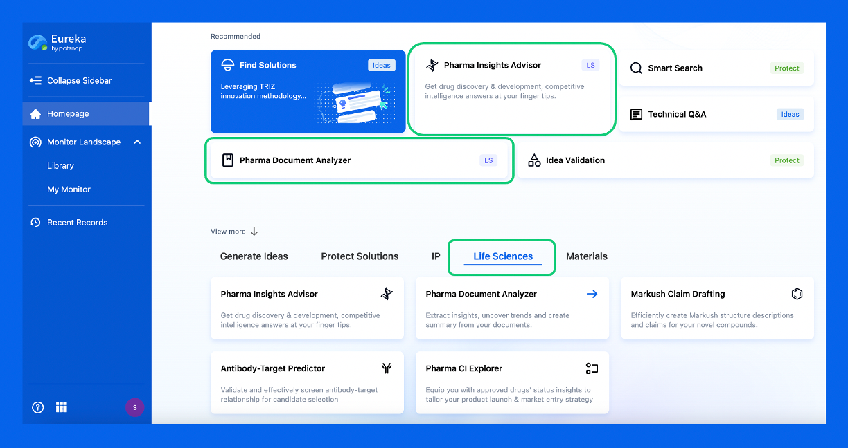
Discover Eureka LS: AI Agents Built for Biopharma Efficiency
Stop wasting time on biopharma busywork. Meet Eureka LS - your AI agent squad for drug discovery.
▶ See how 50+ research teams saved 300+ hours/month
From reducing screening time to simplifying Markush drafting, our AI Agents are ready to deliver immediate value. Explore Eureka LS today and unlock powerful capabilities that help you innovate with confidence.

AI Agents Built for Biopharma Breakthroughs
Accelerate discovery. Empower decisions. Transform outcomes.
Get started for free today!
Accelerate Strategic R&D decision making with Synapse, PatSnap’s AI-powered Connected Innovation Intelligence Platform Built for Life Sciences Professionals.
Start your data trial now!
Synapse data is also accessible to external entities via APIs or data packages. Empower better decisions with the latest in pharmaceutical intelligence.