Request Demo
How to Load a DNA Ladder in Agarose Gel Properly
9 May 2025
Loading a DNA ladder in agarose gel is a fundamental procedure in molecular biology labs, enabling researchers to estimate the size of DNA fragments in their samples. This step-by-step guide will ensure that you load the DNA ladder properly, leading to clear and interpretable results.
Before beginning, gather all the necessary materials: an agarose gel, electrophoresis buffer, DNA ladder, loading dye, micropipette with tips, protective gear, and a gel electrophoresis apparatus. Safety first – always wear gloves and goggles to protect yourself from potential hazards associated with ethidium bromide or other staining agents.
Start by preparing your agarose gel. Depending on the size of the DNA fragments you expect to resolve, choose an appropriate concentration of agarose. For most routine DNA separation, a concentration between 0.8% and 2% is adequate. Melt the agarose in the electrophoresis buffer by heating it in a microwave, and pour it into the gel tray with a comb in place. Allow the gel to set at room temperature until it solidifies, usually around 20 to 30 minutes.
While the gel is setting, prepare the DNA ladder. Mix the ladder with an appropriate volume of loading dye. This dye serves multiple purposes: it adds density to the sample, ensuring it sinks into the wells, and it helps track the progress of the electrophoresis. Check the manufacturer's instructions for the recommended dilution of the ladder with the loading dye.
Once the gel has set, carefully remove the comb, ensuring you do not disturb the wells. Place the gel in the electrophoresis chamber and cover it with running buffer. It is crucial that the gel is completely submerged to allow the current to pass through effectively.
Now it’s time to load the DNA ladder. Using a micropipette, carefully draw up the ladder-dye mixture. Steady your hand and slowly insert the pipette tip just above the bottom of the well, being careful not to puncture the gel. Gently release the solution into the well. Aim to load the ladder in one of the lanes on either edge of the gel to serve as a reference for the samples you will load in the center lanes.
If you are loading multiple samples, it is critical to ensure that the ladder and samples do not overflow into each other’s wells, which can lead to cross-contamination and inaccurate results. Make sure each sample and the ladder have sufficient space and are loaded evenly.
Once all lanes are loaded, secure the lid of the electrophoresis chamber and connect it to a power supply. Run the gel at an appropriate voltage and duration, again referring to the ladder manufacturer’s guidelines. Typically, 5 to 8 volts per centimeter of gel is effective, but this may vary based on the size of the gel and the expected fragment sizes.
During electrophoresis, the loading dye will migrate ahead of the DNA, allowing you to monitor the progress. Once the dye fronts have moved sufficiently (usually about two-thirds of the way down the gel), turn off the power supply.
Carefully remove the gel for visualization. Depending on the stain used, this might involve viewing under UV light if ethidium bromide or another fluorescent dye was used. Compare the bands formed by your DNA samples against the ladder to determine fragment sizes.
In conclusion, loading a DNA ladder properly is crucial for obtaining reliable and interpretable results in gel electrophoresis. By following these steps meticulously, you can ensure accurate size estimation of your DNA fragments, facilitating your molecular biology research.
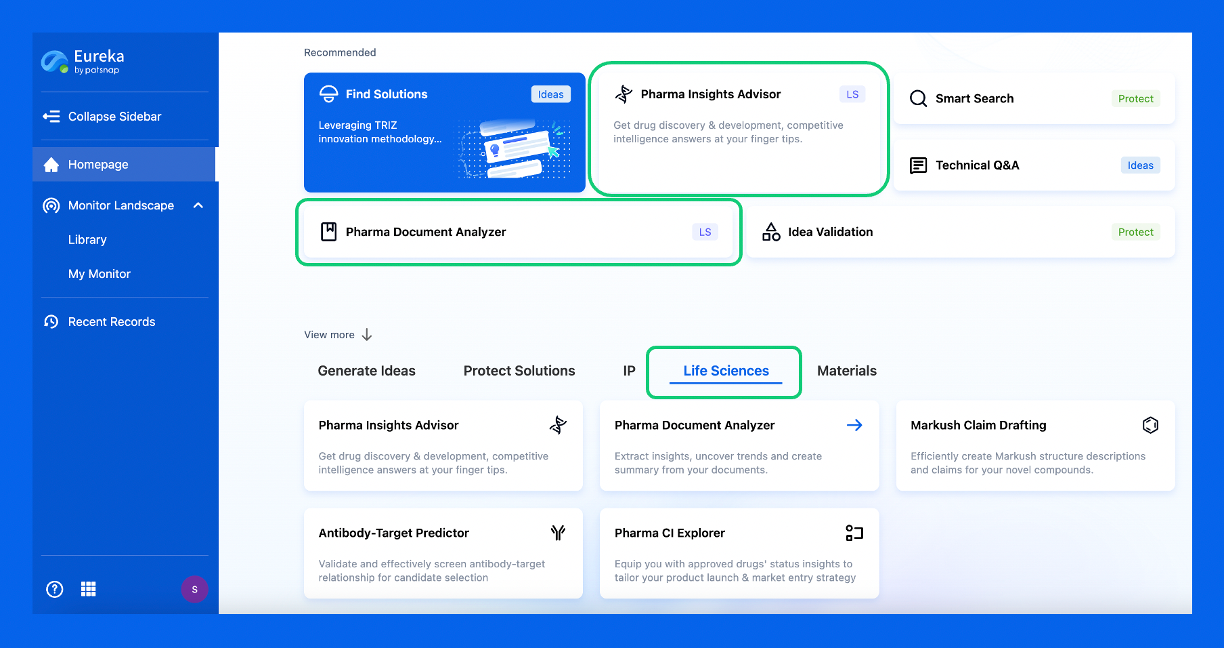
Discover Eureka LS: AI Agents Built for Biopharma Efficiency
Stop wasting time on biopharma busywork. Meet Eureka LS - your AI agent squad for drug discovery.
▶ See how 50+ research teams saved 300+ hours/month
From reducing screening time to simplifying Markush drafting, our AI Agents are ready to deliver immediate value. Explore Eureka LS today and unlock powerful capabilities that help you innovate with confidence.

AI Agents Built for Biopharma Breakthroughs
Accelerate discovery. Empower decisions. Transform outcomes.
Get started for free today!
Accelerate Strategic R&D decision making with Synapse, PatSnap’s AI-powered Connected Innovation Intelligence Platform Built for Life Sciences Professionals.
Start your data trial now!
Synapse data is also accessible to external entities via APIs or data packages. Empower better decisions with the latest in pharmaceutical intelligence.