Request Demo
How to perform a cell fractionation experiment?
27 May 2025
Introduction to Cell Fractionation
Cell fractionation is a vital laboratory technique used to isolate and study the different components within a cell. This method allows researchers to separate cellular organelles, membranes, and other structures, enabling a detailed analysis of their functions and interactions. This article will guide you through the process of performing a cell fractionation experiment, from preparation to analysis.
Preparation and Materials
Before diving into the procedure, it's essential to gather all necessary materials and equipment. You will need:
- A cell culture or tissue sample
- Homogenization buffer (appropriate for the cell type)
- Homogenizer or blender
- Centrifuge with appropriate rotor
- Centrifuge tubes
- Ice and cold storage
- Microscope for analysis
- Assay kits for protein or enzyme activity, if needed
Ensure all equipment is calibrated and that the working area is clean and organized. Keeping the samples cold is crucial to prevent degradation, so have ice or a cold room available.
Step 1: Cell Homogenization
The first step in cell fractionation is homogenization, where cells are broken open to release their contents. The method of homogenization can vary based on the cell type but commonly involves mechanical disruption using a homogenizer or blender. The process should be gentle enough to preserve organelle integrity.
- Place your cell sample in a homogenization buffer that maintains pH and osmotic balance.
- Use the homogenizer to apply sheer force to break open the cells. Perform this step on ice to minimize enzyme activity that could degrade organelles.
- Check under a microscope to ensure cells are adequately broken. You should see released organelles without significant debris.
Step 2: Differential Centrifugation
Differential centrifugation is the primary technique used to separate various cellular components based on their size and density.
- Transfer the homogenate to a centrifuge tube and centrifuge at a low speed (e.g., 600 x g) to pellet the nuclei.
- Carefully collect the supernatant without disturbing the pellet. This supernatant contains other cellular components.
- Centrifuge the supernatant at a higher speed (e.g., 10,000 x g) to pellet mitochondria, lysosomes, and peroxisomes. Collect and label the pellet and supernatant separately.
- Continue increasing centrifugation speeds to further separate smaller components like microsomes and ribosomes. Each speed will yield a different pellet containing specific organelles.
Step 3: Purification and Analysis
Once you have isolated the desired fractions, you may need to perform additional purification steps, such as density gradient centrifugation, to enhance purity.
- For density gradient centrifugation, prepare a gradient (e.g., sucrose gradient) and layer your fraction onto it. Centrifuge at high speed to allow organelles to settle into layers according to density.
- Collect the purified fractions and analyze them using assays or microscopy. Protein assays, enzyme activity measurements, or immunoblotting can validate the presence and purity of specific organelles.
Troubleshooting and Optimization
Like any laboratory technique, cell fractionation may require optimization. Here are some tips to consider:
- Homogenization conditions may need adjustment to ensure cell breakage without damaging organelles—experiment with different shear forces and buffer compositions.
- Ensure centrifugation speeds and times are appropriate for the organelles of interest, as they may vary between cell types.
- Validate the integrity and purity of fractions regularly to ensure reliable results.
Conclusion
Cell fractionation is a powerful technique that offers insights into cellular structure and function. By following a structured approach from preparation through analysis, researchers can effectively isolate and study cellular components. Remember, each cell type may require specific adjustments to the protocol, so flexibility and optimization are key to successful experiments. With a solid understanding of this method, you can explore cellular mysteries at a deeper level, contributing valuable knowledge to the scientific community.
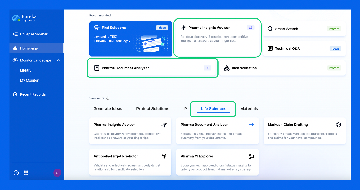
Discover Eureka LS: AI Agents Built for Biopharma Efficiency
Stop wasting time on biopharma busywork. Meet Eureka LS - your AI agent squad for drug discovery.
▶ See how 50+ research teams saved 300+ hours/month
From reducing screening time to simplifying Markush drafting, our AI Agents are ready to deliver immediate value. Explore Eureka LS today and unlock powerful capabilities that help you innovate with confidence.

AI Agents Built for Biopharma Breakthroughs
Accelerate discovery. Empower decisions. Transform outcomes.
Get started for free today!
Accelerate Strategic R&D decision making with Synapse, PatSnap’s AI-powered Connected Innovation Intelligence Platform Built for Life Sciences Professionals.
Start your data trial now!
Synapse data is also accessible to external entities via APIs or data packages. Empower better decisions with the latest in pharmaceutical intelligence.