Request Demo
How to Set Up a CHO Cell Culture Workflow in Your Lab
29 April 2025
Setting up a CHO cell culture workflow in your lab can be an exciting and rewarding endeavor, offering opportunities for various biotechnological applications, including recombinant protein production and therapeutic development. CHO, or Chinese Hamster Ovary, cells are widely used in biological research due to their rapid growth, adaptability to various culture conditions, and ability to produce high-quality proteins. This guide will walk you through the essential steps to establish a successful CHO cell culture process in your laboratory.
The first step in setting up your CHO cell culture workflow is to ensure that you have the necessary equipment and materials. This includes a sterile workspace, usually a biological safety cabinet, to prevent contamination. You'll also need a CO2 incubator to maintain the appropriate temperature and pH levels for cell growth. Other essential materials include culture media, such as Ham's F-12 or DMEM/F-12, supplemented with fetal bovine serum (FBS) or other growth factors specific to your cell line's requirements. Additionally, you'll need culture vessels, such as flasks or multi-well plates, as well as pipettes, sterile pipette tips, and centrifuge tubes.
Once your workspace is set up and your materials are ready, the next step is to thaw and revive your CHO cells. Start by retrieving a vial of frozen CHO cells from liquid nitrogen storage. Carefully thaw the vial in a 37°C water bath while gently swirling it to ensure even heating. Once thawed, immediately transfer the cells to a sterile centrifuge tube containing pre-warmed culture media. Centrifuge the cells at a low speed to pellet them, then carefully remove the supernatant; this step helps remove residual DMSO from the freezing media. Gently resuspend the cell pellet in fresh culture media and transfer the cells to a culture flask. Place the flask in the CO2 incubator, ensuring it is set to the appropriate temperature (usually 37°C) and CO2 concentration (typically 5%).
Monitoring and maintaining your CHO cell culture is crucial for optimal growth and viability. Regularly check the cells under a microscope to assess confluency, morphology, and any signs of contamination. Change the culture media every two to three days or when the media becomes yellow, indicating a drop in pH. To split confluent cultures, remove the spent media and wash the cells with a sterile phosphate-buffered saline (PBS) solution. Add a trypsin-EDTA solution to detach the cells from the surface, incubate for a few minutes, then neutralize the trypsin with fresh media. Gently pipette the cell suspension to ensure single-cell dispersal, and seed the cells into new culture vessels at the desired density.
Scaling up your CHO cell culture for larger experiments or protein production can be achieved by transferring cells to larger flasks or bioreactors. When scaling up, ensure that all equipment is appropriately sized and that the culture conditions remain consistent to support healthy cell growth.
Quality control is vital in a CHO cell culture workflow. Periodically test for mycoplasma contamination and verify cell identity using genetic or protein-based assays. Regularly review cell growth curves and protein expression levels to ensure that your cultures are performing as expected.
By following these guidelines and maintaining a sterile and well-organized lab environment, you can successfully set up and manage a CHO cell culture workflow. With practice, you'll be able to optimize conditions specifically for your research needs, contributing to meaningful advancements in biotechnology and therapeutic development.
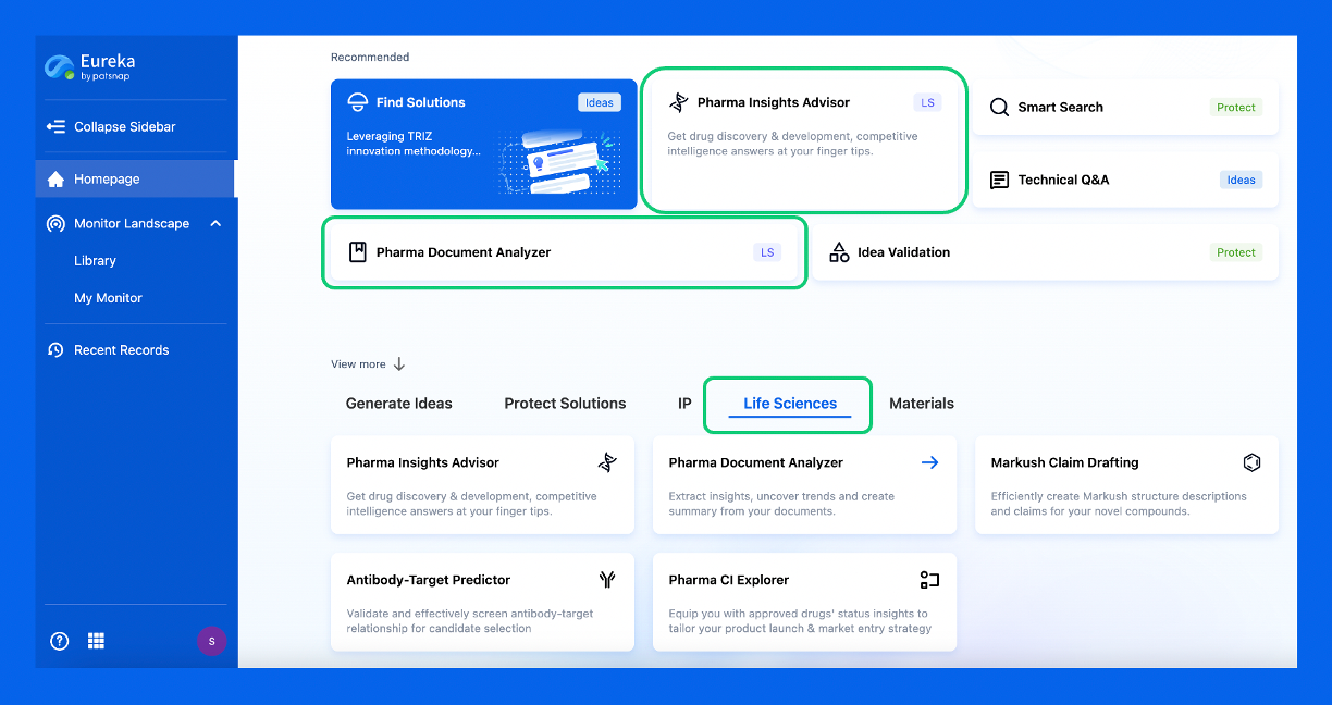
Discover Eureka LS: AI Agents Built for Biopharma Efficiency
Stop wasting time on biopharma busywork. Meet Eureka LS - your AI agent squad for drug discovery.
▶ See how 50+ research teams saved 300+ hours/month
From reducing screening time to simplifying Markush drafting, our AI Agents are ready to deliver immediate value. Explore Eureka LS today and unlock powerful capabilities that help you innovate with confidence.

AI Agents Built for Biopharma Breakthroughs
Accelerate discovery. Empower decisions. Transform outcomes.
Get started for free today!
Accelerate Strategic R&D decision making with Synapse, PatSnap’s AI-powered Connected Innovation Intelligence Platform Built for Life Sciences Professionals.
Start your data trial now!
Synapse data is also accessible to external entities via APIs or data packages. Empower better decisions with the latest in pharmaceutical intelligence.