Request Demo
Protein Quantification Protocol: From Sample Preparation to Data Analysis
9 May 2025
Protein quantification is a fundamental process in the field of biochemistry and molecular biology, serving as a critical step in various research and clinical applications. Understanding the amount of protein present in a sample is vital for downstream analyses such as enzyme activity assays, Western blotting, and mass spectrometry. This blog outlines a comprehensive protein quantification protocol, detailing the journey from sample preparation to data analysis.
The initial phase of protein quantification begins with sample preparation. This stage is crucial since the accuracy and reliability of the quantification depend significantly on the quality of the sample. Proteins must be extracted efficiently while minimizing degradation and contamination. Depending on the source, whether it be tissue, cell lysate, or a purified sample, the methods may vary. Typically, proteins are extracted using a buffer solution that maintains pH and ionic strength to preserve protein integrity. The inclusion of protease inhibitors is often necessary to prevent proteolytic degradation. Following extraction, the sample is usually clarified by centrifugation to remove debris and insoluble materials.
Once the sample is ready, it is essential to select an appropriate protein quantification method. Several techniques are commonly employed, each with its unique advantages and limitations. The Bradford assay, for example, is one of the most widely used methods due to its simplicity and speed. It relies on the binding of Coomassie Brilliant Blue dye to proteins, resulting in a color change proportional to the protein concentration. Despite its popularity, the Bradford assay is sensitive to interference from detergents and other chemicals commonly found in lysis buffers.
Alternatively, the Bicinchoninic Acid (BCA) assay is favored for its compatibility with a broader range of substances, including detergents. The BCA assay involves the reduction of Cu^2+ ions to Cu^1+ by proteins in an alkaline environment, followed by the formation of a purple-colored complex with bicinchoninic acid. This method is more tolerant of common contaminants but requires a longer incubation period compared to the Bradford assay.
For researchers requiring high sensitivity and precision, the Lowry method remains a gold standard. This approach involves a reaction between protein and an alkaline copper tartrate solution, followed by the addition of the Folin-Ciocalteu reagent, resulting in a blue color change. Although this method is more time-consuming and sensitive to interfering substances, it provides reliable and accurate results, particularly for samples with low protein concentration.
After selecting a suitable quantification method, the next step is to perform the assay and generate data. Standard curves are essential for quantification, as they allow for the determination of protein concentration in unknown samples. A series of known protein standards are assayed alongside the samples, and their absorbance is measured using a spectrophotometer. The resulting absorbance values are plotted to generate a standard curve, which is then used to interpolate the protein concentration of the unknown samples.
Data analysis in protein quantification involves interpreting the results and ensuring their validity. It is crucial to confirm that the samples fall within the linear range of the standard curve to ensure accurate quantification. Researchers must also account for any potential sources of error, such as pipetting inaccuracies or sample contamination. Statistical analysis may be applied to assess the reproducibility of the measurements and to determine the significance of differences between experimental conditions.
In conclusion, protein quantification is a multi-step process requiring careful attention to sample preparation, method selection, and data analysis. By following a systematic protocol, researchers can obtain reliable and accurate protein measurements, providing a solid foundation for further biochemical investigations. Whether you are quantifying proteins for basic research, clinical diagnostics, or biotechnological applications, understanding and executing each step of the protocol with precision is key to achieving meaningful results.
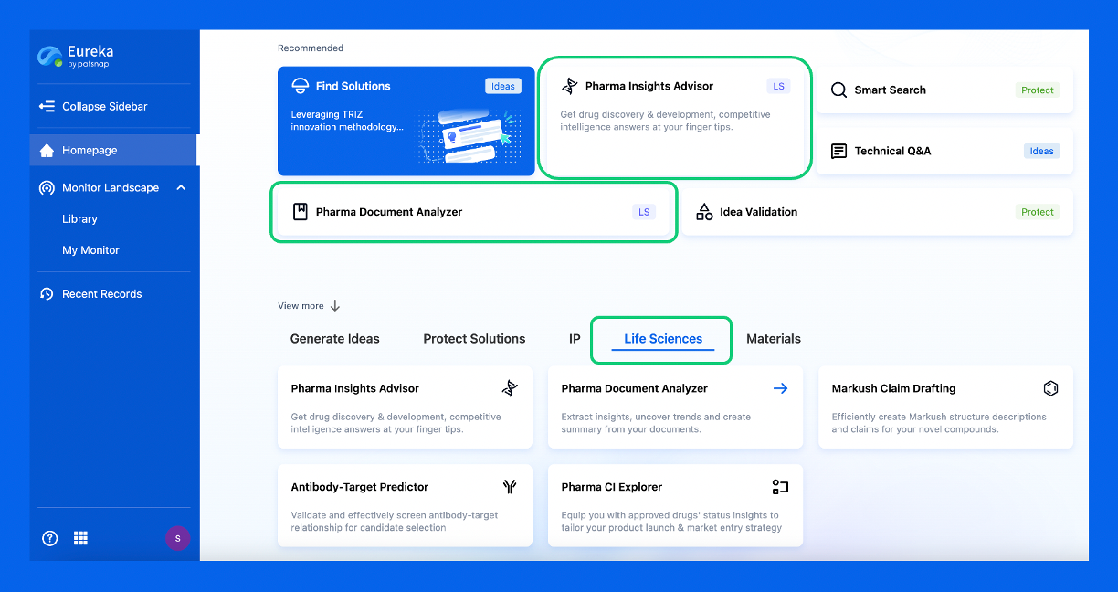
Discover Eureka LS: AI Agents Built for Biopharma Efficiency
Stop wasting time on biopharma busywork. Meet Eureka LS - your AI agent squad for drug discovery.
▶ See how 50+ research teams saved 300+ hours/month
From reducing screening time to simplifying Markush drafting, our AI Agents are ready to deliver immediate value. Explore Eureka LS today and unlock powerful capabilities that help you innovate with confidence.

AI Agents Built for Biopharma Breakthroughs
Accelerate discovery. Empower decisions. Transform outcomes.
Get started for free today!
Accelerate Strategic R&D decision making with Synapse, PatSnap’s AI-powered Connected Innovation Intelligence Platform Built for Life Sciences Professionals.
Start your data trial now!
Synapse data is also accessible to external entities via APIs or data packages. Empower better decisions with the latest in pharmaceutical intelligence.