Request Demo
Western Blot Step-by-Step: From Sample Prep to Band Detection
29 April 2025
Western blotting is a powerful and widely used technique in molecular biology and biochemistry to detect specific proteins in a sample. This method involves several key steps, each crucial for ensuring accurate and reliable results. Below is a detailed walkthrough of the Western blotting process, from sample preparation to band detection.
The first step in Western blotting is sample preparation. This involves lysing cells or tissues to extract proteins. It's essential to use an appropriate lysis buffer that contains detergents like SDS or Triton X-100, protease inhibitors, and possibly phosphatase inhibitors, depending on the target protein. The lysate is then centrifuged to remove debris, and the supernatant, which contains the proteins, is collected. Protein concentration should be determined using assays such as the Bradford, Bicinchoninic Acid (BCA), or Lowry methods to ensure equal loading across samples.
Once the proteins are prepared, they need to be separated by size through SDS-PAGE (sodium dodecyl sulfate-polyacrylamide gel electrophoresis). SDS is an anionic detergent that denatures proteins and gives them a uniform negative charge, allowing separation based solely on molecular weight. Samples are mixed with a loading buffer containing SDS and a reducing agent such as beta-mercaptoethanol or dithiothreitol (DTT), then heated to denature the proteins completely. They are loaded into the wells of a polyacrylamide gel and subjected to an electric field, which causes the proteins to migrate through the gel matrix. Smaller proteins move faster and thus travel further than larger proteins.
After electrophoresis, the proteins are transferred from the gel to a membrane, typically made of nitrocellulose or polyvinylidene difluoride (PVDF). This step, known as the blotting step, is crucial because membranes provide a solid support that can be easily probed with antibodies. The transfer is generally performed using an electric field in a wet or semi-dry transfer system. Ensuring efficient transfer is vital; thus, factors such as the time, voltage, and buffer composition need to be optimized.
Following transfer, it's essential to block the membrane to prevent non-specific binding of antibodies. This is achieved by incubating the membrane in a blocking buffer, typically containing 5% non-fat milk or BSA in Tris-buffered saline with Tween 20 (TBST). Blocking reduces background noise and enhances the specificity of antibody binding.
The next step is antibody probing, where the membrane is incubated with a primary antibody specific to the target protein. This antibody binds to its target, and after thorough washing to remove unbound antibodies, a secondary antibody is applied. The secondary antibody is typically conjugated to an enzyme like horseradish peroxidase (HRP) or alkaline phosphatase. It binds to the primary antibody and is used to amplify the signal.
Detection of the target protein is accomplished by adding a substrate that reacts with the enzyme conjugated to the secondary antibody, producing a detectable signal. For HRP, chemiluminescent substrates are commonly used, whereby the enzyme catalyzes a reaction that emits light, captured using film or a digital imaging system. For alkaline phosphatase, chromogenic substrates can produce a colored precipitate visible on the membrane.
Finally, the signal is quantified, often by densitometry, to determine the relative abundance of the protein. Proper controls, including loading controls like housekeeping proteins (e.g., actin or tubulin), are essential for accurate interpretation of the results.
In conclusion, Western blotting is a meticulous process requiring careful attention to detail at each step. From effective sample preparation and careful electrophoretic separation to precise transfer, blocking, and detection, each stage is crucial for obtaining reliable and interpretable results. By following this step-by-step guide, researchers can achieve accurate protein detection and quantification, advancing their understanding of protein expression and function.
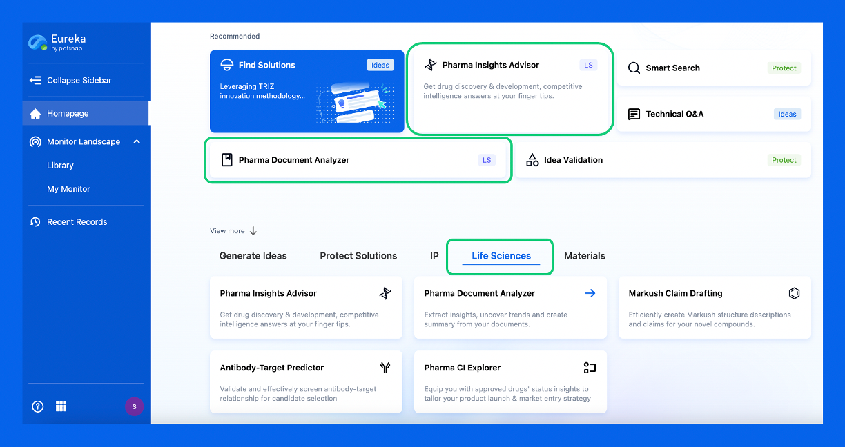
Discover Eureka LS: AI Agents Built for Biopharma Efficiency
Stop wasting time on biopharma busywork. Meet Eureka LS - your AI agent squad for drug discovery.
▶ See how 50+ research teams saved 300+ hours/month
From reducing screening time to simplifying Markush drafting, our AI Agents are ready to deliver immediate value. Explore Eureka LS today and unlock powerful capabilities that help you innovate with confidence.

AI Agents Built for Biopharma Breakthroughs
Accelerate discovery. Empower decisions. Transform outcomes.
Get started for free today!
Accelerate Strategic R&D decision making with Synapse, PatSnap’s AI-powered Connected Innovation Intelligence Platform Built for Life Sciences Professionals.
Start your data trial now!
Synapse data is also accessible to external entities via APIs or data packages. Empower better decisions with the latest in pharmaceutical intelligence.