What Is Western Blot? A Step-by-Step Guide to Protein Detection
Western blotting is a widely used analytical technique in molecular biology and biochemistry for detecting specific proteins in a sample. This method is invaluable for researchers seeking to understand protein expression, identify specific proteins, and diagnose diseases. This guide will walk you through the step-by-step process of Western blotting, explaining each stage in detail.
The process begins with sample preparation. Proper sample preparation is crucial as it ensures accurate results. Typically, proteins are extracted from cells or tissues using a lysis buffer, which breaks down cell membranes and releases proteins. It's important to keep the samples on ice to prevent protein degradation. Protease inhibitors are often added to the lysis buffer to protect the proteins from being broken down by enzymes.
After obtaining the protein extract, the next step is protein quantification. Accurate measurement of protein concentration is necessary to ensure equal loading of samples across the gel. This is commonly done using colorimetric assays, such as the Bradford or BCA assay, which provide a reliable estimate of protein concentration.
The prepared samples are then mixed with a loading buffer that contains SDS (sodium dodecyl sulfate), a detergent that denatures proteins and imparts a negative charge. This ensures that the proteins are separated based solely on their size and not their charge or shape. Heating the samples helps to ensure complete denaturation of the proteins.
Once the samples are ready, they are loaded onto a polyacrylamide gel for electrophoresis. The gel consists of a stacking gel and a resolving gel. The stacking gel, with its lower pH, concentrates the proteins into thin bands, while the resolving gel, with its higher pH, separates the proteins based on their molecular weight. As an electric current is applied, proteins migrate through the gel, with smaller proteins moving faster than larger ones.
After electrophoresis, proteins are transferred from the gel onto a membrane, typically made of nitrocellulose or PVDF (polyvinylidene fluoride). This step, known as blotting, is crucial for immobilizing the proteins so they can be probed with antibodies. The transfer can be accomplished using either a wet or semi-dry transfer system, with the choice depending on the specific requirements of the experiment.
Blocking the membrane is the next step, essential to prevent nonspecific binding of antibodies. This is usually done by incubating the membrane in a solution containing milk or BSA (bovine serum albumin). These proteins coat the membrane, ensuring that antibodies bind only to the target proteins.
Following blocking, the membrane is incubated with a primary antibody specific to the protein of interest. This antibody binds to the target protein, forming a complex that is crucial for detection. After washing away unbound primary antibodies, a secondary antibody is added. This antibody is conjugated to an enzyme, such as horseradish peroxidase (HRP) or alkaline phosphatase, which will facilitate detection.
Finally, the presence of the target protein is revealed through a detection method. When the enzyme on the secondary antibody reacts with its substrate, it produces a signal – often a colored precipitate or chemiluminescent light – that can be captured using imaging systems or X-ray film. The intensity of the signal correlates with the amount of protein, allowing for both qualitative and quantitative analysis.
Western blotting is a powerful tool for protein analysis, offering high specificity and sensitivity. Each step, from sample preparation to detection, is vital for obtaining accurate and reliable results. With practice and attention to detail, researchers can effectively use Western blotting to uncover valuable insights into protein function and expression.
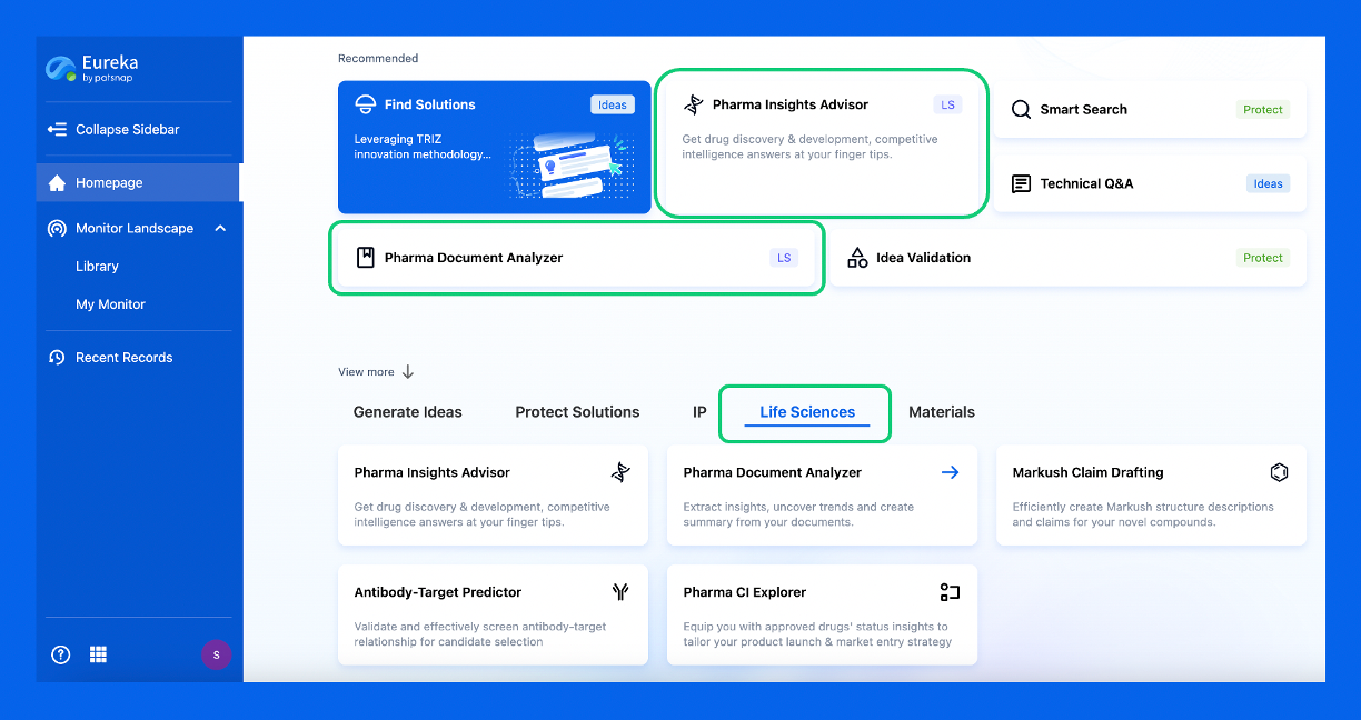
Discover Eureka LS: AI Agents Built for Biopharma Efficiency
Stop wasting time on biopharma busywork. Meet Eureka LS - your AI agent squad for drug discovery.
▶ See how 50+ research teams saved 300+ hours/month
From reducing screening time to simplifying Markush drafting, our AI Agents are ready to deliver immediate value. Explore Eureka LS today and unlock powerful capabilities that help you innovate with confidence.
