Request Demo
Last update 08 May 2025
5D-4
Last update 08 May 2025
Overview
Basic Info
Drug Type Small molecule drug |
Synonyms |
Target |
Action inhibitors |
Mechanism TOPBP1 inhibitors(DNA topoisomerase II binding protein 1 inhibitors) |
Therapeutic Areas |
Active Indication |
Inactive Indication- |
Originator Organization |
Active Organization |
Inactive Organization- |
License Organization- |
Drug Highest PhasePreclinical |
First Approval Date- |
Regulation- |

Related
100 Clinical Results associated with 5D-4
Login to view more data
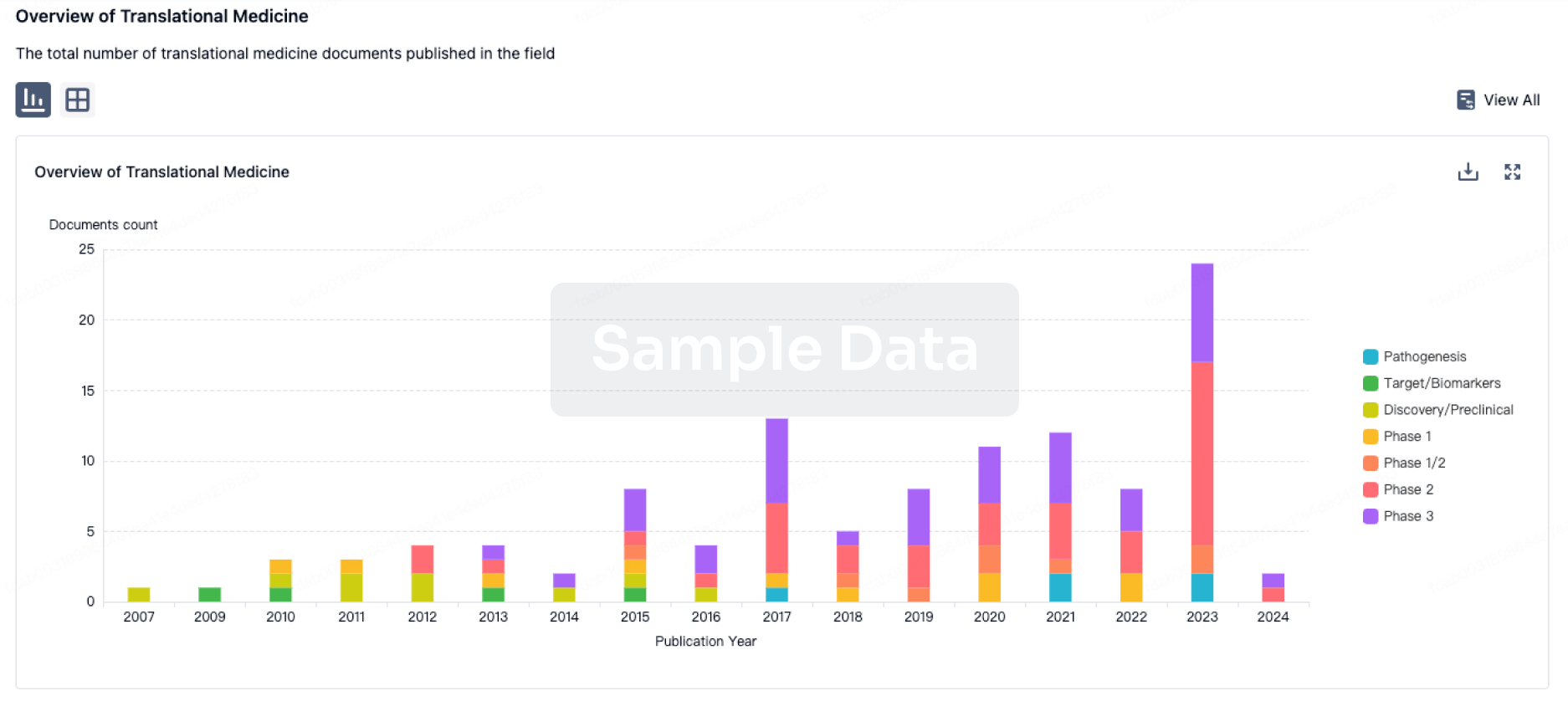
100 Translational Medicine associated with 5D-4
Login to view more data
100 Patents (Medical) associated with 5D-4
Login to view more data
25
Literatures (Medical) associated with 5D-401 May 2025Pathology - Research and Practice
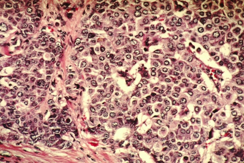
Intracytoplasmic accumulation of keratan sulfate is a hallmark of granular cell tumor
Article
Author: Yonemoto, Natsumi ; Yoshida, Hisato ; Fukushima, Mana ; Kobayashi, Motohiro ; Akama, Tomoya O ; Hoshino, Hitomi ; Muramoto, Akifumi
01 Jun 2024Laboratory Investigation
Structural Elucidation and Prognostic Relevance of 297-11A-Sulfated Glycans in Ovarian Carcinoma
Article
Author: Khoo, Kay-Hooi ; Kobayashi, Motohiro ; Fukushima, Mana ; Hoshino, Hitomi ; Kogami, Akiya ; Yamamoto, Makoto ; Chen, Ya-Ying ; Inoue, Daisuke ; Akama, Tomoya O ; Yoshida, Yoshio
31 Oct 2023Proceedings of the National Academy of Sciences of the United States of AmericaQ1 · CROSS-FIELD
A small-molecule inhibitor of TopBP1 exerts anti-MYC activity and synergy with PARP inhibitors.
Q1 · CROSS-FIELD
Article
Author: Song, Yongcheng ; Liu, Kang ; Lin, Weei-Chin ; Folly-Kossi, Helena ; Lin, Fang-Tsyr ; Lin, Shwu-Jiuan ; Garan, Lidija A Wilhelms
1
News (Medical) associated with 5D-427 Oct 2023
Gene TherapyClinical Result
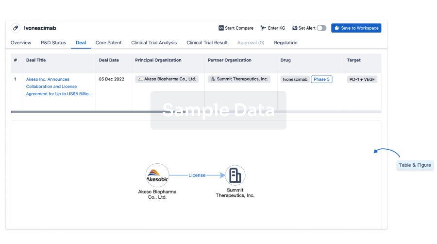
100 Deals associated with 5D-4
Login to view more data
R&D Status
10 top R&D records. to view more data
Login
| Indication | Highest Phase | Country/Location | Organization | Date |
|---|---|---|---|---|
| Breast Cancer | Preclinical | United States | 31 Oct 2023 | |
| Ovarian Cancer | Preclinical | United States | 31 Oct 2023 |
Login to view more data
Clinical Result
Clinical Result
Indication
Phase
Evaluation
View All Results
Login to view more data
Translational Medicine
Boost your research with our translational medicine data.
login
or
Deal
Boost your decision using our deal data.
login
or
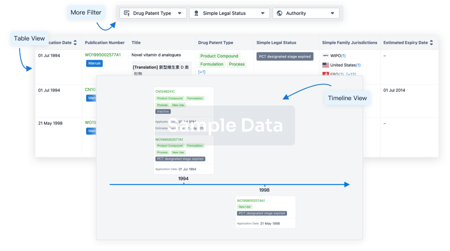
Core Patent
Boost your research with our Core Patent data.
login
or
Clinical Trial
Identify the latest clinical trials across global registries.
login
or
Approval
Accelerate your research with the latest regulatory approval information.
login
or
Regulation
Understand key drug designations in just a few clicks with Synapse.
login
or
AI Agents Built for Biopharma Breakthroughs
Accelerate discovery. Empower decisions. Transform outcomes.
Get started for free today!
Accelerate Strategic R&D decision making with Synapse, PatSnap’s AI-powered Connected Innovation Intelligence Platform Built for Life Sciences Professionals.
Start your data trial now!
Synapse data is also accessible to external entities via APIs or data packages. Empower better decisions with the latest in pharmaceutical intelligence.
Bio
Bio Sequences Search & Analysis
Sign up for free
Chemical
Chemical Structures Search & Analysis
Sign up for free