Request Demo
Last update 13 Dec 2025
COTI-2
Last update 13 Dec 2025
Overview
Basic Info
Drug Type Small molecule drug |
Synonyms COTI 2 |
Target |
Action stimulants |
Mechanism p53 stimulants(Tumor protein p53 stimulants) |
Therapeutic Areas |
Active Indication- |
Inactive Indication |
Originator Organization |
Active Organization- |
Inactive Organization |
License Organization- |
Drug Highest PhasePendingPhase 1 |
First Approval Date- |
Regulation- |
Login to view timeline
Structure/Sequence
Molecular FormulaC19H22N6S |
InChIKeyUTDAKQMBNSHJJB-UHFFFAOYSA-N |
CAS Registry1039455-84-9 |
Related
1
Clinical Trials associated with COTI-2NCT02433626
A Phase 1 Study of COTI-2 as Monotherapy or Combination Therapy for the Treatment of Advanced or Recurrent Malignancies
Activity of COTI-2 has been demonstrated in various cancer tumor models. With its p53- and AKT-based mechanisms of action, COTI-2 is anticipated to be highly relevant in treatment of patients with gynecologic malignancies or head and neck squamous cell carcinoma (HNSCC) as well as a variety of other tumor types.
This study is designed primarily to assess the safety and tolerability of COTI-2 monotherapy or combination therapy in patients with advanced and recurrent malignancies to establish a recommended Phase 2 dose (RP2D) for future studies.
Patients are currently being recruited for Part 3 of the study.
Critical Outcome Technologies Inc. has been renamed to Cotinga Pharmaceuticals.
Start Date01 Dec 2015 |
Sponsor / Collaborator |
100 Clinical Results associated with COTI-2
Login to view more data
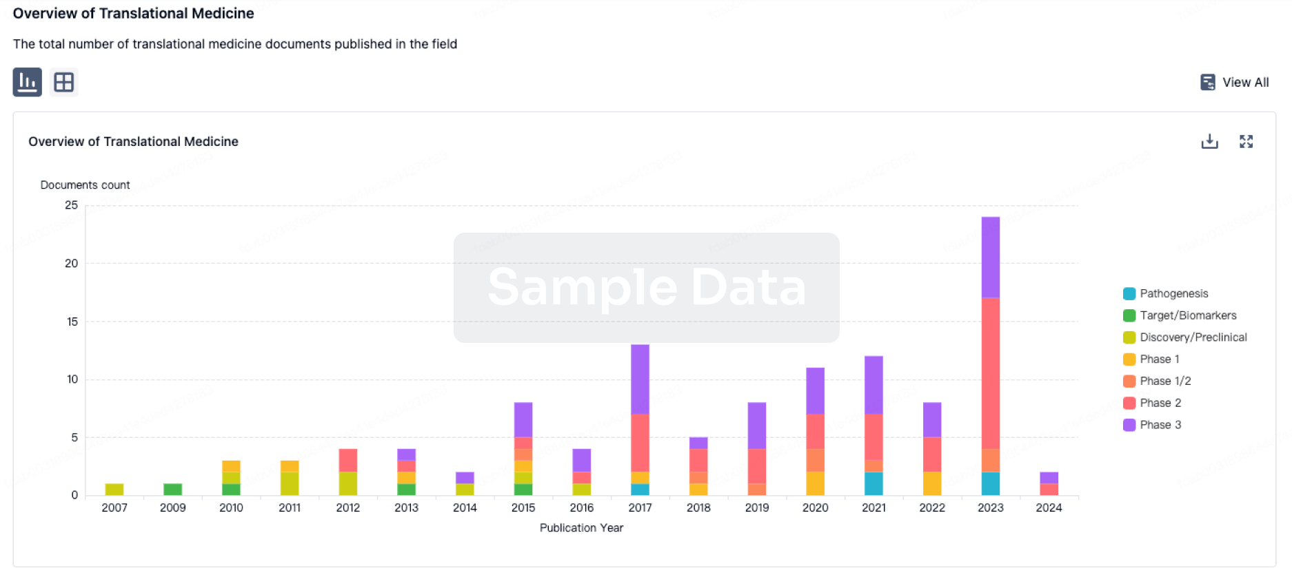
100 Translational Medicine associated with COTI-2
Login to view more data
100 Patents (Medical) associated with COTI-2
Login to view more data
32
Literatures (Medical) associated with COTI-212 Aug 2025·Zhongguo zhen jiu = Chinese acupuncture & moxibustion
[Mechanism of acupuncture on cerebral ischemia-reperfusion injury via p53/SLC7A11/GPX4 signaling pathway in rat models].
Article
Author: Wang, Qi ; Chen, Xia ; Hou, Ziwen ; Wei, Dan ; Kong, Qingjie ; Liu, Yaoyao
Objective:
To explore the neuroprotective effect and underlying mechanism of Xingnao Kaiqiao acupuncture (acupuncture for regaining consciousness and opening orifices) in the rat models of cerebral ischemia-reperfusion injury (CIRI) based on the p53 protein (p53)/solute carrier family 7 member 11 (SLC7A11)/glutathione peroxidase 4 (GPX4) signaling pathway.
Methods:
Of 102 male Wistar rats, 20 rats were randomly collected as a sham-operation group. Using a modified external carotid artery filament insertion method, CIRI models were prepared by occluding the middle cerebral artery in the rest rats. After modeling and excluding 1 non-successfully modeled rat and 1 dead one, the other modeled rats were randomized into a model group, an agonist group, an acupuncture group, and an acupuncture + agonist group, 20 rats in each one. Xingnao Kaiqiao acupuncture therapy was delivered in the rats of the acupuncture group and the acupuncture + agonist group. The acupoints included "Shuigou" (GV26), bilateral "Neiguan" (PC6), and "Sanyinjiao" (SP6) on the affected side. Electroacupuncture was attached to "Neiguan" (PC6) and "Sanyinjiao" (SP6) on the affected side, with dense-disperse wave, a frequency of 2 Hz/15 Hz and intensity of 1 mA. The intervention was delivered twice daily, 20 min each time and for 7 consecutive days. In the agonist group and acupuncture+agonist group, p53 agonist, COTI-2 was intraperitoneally injected (15 mg/kg), once daily for 7 consecutive days. Neurological deficit was evaluated using Zausinger's six-point scale. Cerebral infarction volume was quantified by triphenyl tetrazolium chloride (TTC) staining. Histopathological changes were observed using hematoxylin-eosin (HE) staining. Iron deposition was assessed by Prussian blue staining. Mitochondrial ultrastructure in the ischemic cortex was examined under transmission electron microscopy (TEM). Serum iron (Fe2+) was measured with chromometry. Malondialdehyde (MDA) and glutathione (GSH) levels in the ischemic hippocampus were determined using thiobarbituric acid and microplate assays, respectively. The mean fluorescence intensity of reactive oxygen species (ROS) in the ischemic cortex was analyzed by flow cytometry. The mRNA and protein expression of GPX4, SLC7A11, and p53 in the ischemic hippocampus were evaluated using quantitative real-time PCR (qRT-PCR) and Western blotting, respectively.
Results:
Compared with the sham-operated group, the model group exhibited the decrease in neurological deficit score (P<0.01), and the increase in cerebral infarction volume percentage (P<0.01). The changes of brain tissue were presented in extensive cellular necrosis, pyknotic and deeply-stained nuclei, and vacuolar degeneration. The iron deposition was elevated in cortex and hippocampus (P<0.01), mitochondrial membrane density increased, the cristae was broken or reduced, and the outer membrane ruptured. The levels of Fe2+ and MDA, as well as the mean flourscence intensity of ROS were elevated (P<0.01) and the level of GSH was reduced (P<0.01). The mRNA and protein expression of GPX4 and SLC7A11 was reduced (P<0.01), while that of p53 rose (P<0.01). When compared with the model group, in the agonist group, the neurological deficit score was reduced (P<0.05), the percentage of infarction volume was higher (P<0.01), the histopathological damage was further exacerbated, and the percentage of iron deposition increased in the cortex and hippocampus (P<0.01). The mitochondrial quantity decreased, the membrane density increased, the mitochondrial cristae were broken or reduced, and the outer membrane was ruptured. The levels of Fe2+ and MDA, as well as the mean flourscence intensity of ROS were higher (P<0.01, P<0.05) and the level of GSH was reduced (P<0.05). The mRNA and protein expression of GPX4 and SLC7A11 decreased (P<0.01, P<0.05), while that of p53 was elevated (P<0.01). Besides, in comparison with the model group, the neurological deficit score was higher in the acupuncture group and the acupuncture + agonist group (P<0.01, P<0.05), the percentage of cerebral infarction volume was lower in the acupuncture group (P<0.01), the pathological damage of brain tissue was alleviated in the acupuncture group and the acupuncture + agonist group, and the percentage of iron depositiondecreased in the cortex and hippocampus (P<0.01). The mitochondrial structure was relatively clear, the mitochondrial cristae were fractured or reduced mildly in the acupuncture group and the acupuncture + agonist group. The levels of Fe2+ and MDA, as well as the mean flourscence intensity of ROS were lower (P<0.01) and the level of GSH was higher (P<0.01) in the acupuncture group. The mean fluorescence intensity of ROS were dropped (P<0.01) in the acupuncture + agonist group. The mRNA expression of GPX4 and SLC7A11 was elevated (P<0.01) and that of p53 was reduced (P<0.01, P<0.05) in either the acupuncture group or the acupuncture + agonist group; the protein expression of GPX4 and SLC7A11 rose (P<0.05, P<0.01) and that of p53 was dropped (P<0.01) in the acupuncture group; and the protein expression of p53 was also lower in the acupuncture + agonist group (P<0.05). When compared with the agonist group, in the acupuncture + agonist group, neurological deficit score increased (P<0.01), the percentage of cerebral infarction volume decreased (P<0.01), the pathological brain tissue damage was reduced, the percentage of iron deposition in cortex and hippocampus decreased (P<0.01), the mitochondrial structure was relatively clear and the cristae broken or reduced slightly; the levels of Fe2+ and MDA, as well as the mean fluorescence intensity of ROS were dropped (P<0.01), while the level of GSH increased (P<0.05); the mRNA and protein expression of GPX4 and SLC7411 was elevated (P<0.01, P<0.05), and that of p53 reduced (P<0.01). In comparison with the acupuncture + agonist group, in the acupuncture group, the neurological deficit score increased (P<0.05), the percentage of cerebral infarction volume decreased (P<0.05), the pathological brain tissue damage was alleviated, the percentage of iron deposition in cortex and hippocampus decreased (P<0.01), the mitochondrial structure was normal in tendency; the levels of Fe2+ and MDA, as well as the mean fluorescence intensity of ROS were reduced (P<0.05), while the level of GSH rose (P<0.01); the mRNA and protein expression of GPX4 and SLC7411 was elevated (P<0.01, P<0.05), and that of p53 reduced (P<0.01, P<0.05).
Conclusion:
Xingnao Kaiqiao acupuncture can alleviate neurological damage in CIRI rats, which is obtained probably by inhibiting ferroptosis through p53/SLC7A11/GPX4 pathway.
25 Jul 2024·JOURNAL OF MEDICINAL CHEMISTRY
Isosteric Replacement of Sulfur to Selenium in a Thiosemicarbazone: Promotion of Zn(II) Complex Dissociation and Transmetalation to Augment Anticancer Efficacy
Article
Author: Suleymanoglu, Mediha ; Wijesinghe, Tharushi P. ; Harmer, Jeffrey R. ; Richardson, Vera ; Dharmasivam, Mahendiran ; Richardson, Des R. ; Zhao, Xiao ; Kaya, Busra ; Gholam Azad, Mahan ; Bernhardt, Paul V.
We implemented isosteric replacement of sulfur to selenium in a novel thiosemicarbazone (PPTP4c4mT) to create a selenosemicarbazone (PPTP4c4mSe) that demonstrates potentiated anticancer efficacy and selectivity. Their design specifically incorporated cyclohexyl and styryl moieties to sterically inhibit the approach of their Fe(III) complexes to the oxy-myoglobin heme plane. Importantly, in contrast to the Fe(III) complexes of the clinically trialed thiosemicarbazones Triapine, COTI-2, and DpC, the Fe(III) complexes of PPTP4c4mT and PPTP4c4mSe did not induce detrimental oxy-myoglobin oxidation. Furthermore, PPTP4c4mSe demonstrated more potent antiproliferative activity than the homologous thiosemicarbazone, PPTP4c4mT, with their selectivity being superior or similar, respectively, to the clinically trialed thiosemicarbazone, COTI-2. An advantageous property of the selenosemicarbazone Zn(II) complexes relative to their thiosemicarbazone analogues was their greater transmetalation to Cu(II) complexes in lysosomes. This latter effect probably promoted their antiproliferative activity. Both ligands down-regulated multiple key receptors that display inter-receptor cooperation that leads to aggressive and resistant breast cancer.
25 Apr 2024·Journal of medicinal chemistry
Correction to “Steric Blockade of Oxy-Myoglobin Oxidation by Thiosemicarbazones: Structure–Activity Relationships of the Novel PPP4pT Series’’
Author: Gholam Azad, Mahan ; Gonzalvez, Miguel A. ; Harmer, Jeffrey R. ; Wijesinghe, Tharushi P. ; Richardson, Des R. ; Kaya, Busra ; Bernhardt, Paul V. ; Dharmasivam, Mahendiran
The di-2-pyridylketone thiosemicarbazones demonstrated marked anticancer efficacy, prompting progression of DpC to clin. trials.However, DpC induced deleterious oxy-myoglobin oxidation, stifling development.To address this, novel substituted Ph thiosemicarbazone (PPP 4pT) analogs and their Fe(III), Cu(II), and Zn(II) complexes were preparedThe PPP 4pT analogs demonstrated potent antiproliferative activity (IC50: 0.009-0.066μM), with the 1:1 Cu:L complexes showing the greatest efficacy.Substitutions leading to decreased redox potential of the PPP 4pT:Cu(II) complexes were associated with higher antiproliferative activity, while increasing potential correlated with increased redox activity.Surprisingly, there was no correlation between redox activity and antiproliferative efficacy.The PPP 4pT:Fe(III) complexes attenuated oxy-myoglobin oxidation significantly more than the clin. trialed thiosemicarbazones, Triapine, COTI-2, and DpC, or earlier thiosemicarbazone series.Incorporation of phenyl- and styryl-substituents led to steric blockade, preventing approach of the PPP 4pT:Fe(III) complexes to the heme plane and its oxidationThe 1:1 Cu(II):PPP 4pT complexes were inert to transmetalation and did not induce oxy-myoglobin oxidation
2
News (Medical) associated with COTI-223 Jan 2017
Summary
Critical Outcome Technologies Inc COTI is a biopharmaceutical company that develops targeted therapeutics with focus on oncology. Its lead program COTI2 is a novel small molecule activator of misfolded mutant p53 proteins for the treatment of ovarian and other gynecological cancers. The company's other pipeline products include COTI219, COTI4, and COTI58 for small cell lung cancer; and other products for Alzheimer's disease, HIV integrase inhibitors, and multiple sclerosis. COTI uses its proprietary technology Chemsas, a multistage computational platform technology based on hybrid of machine learning technologies and proprietary algorithms which allows accurate prediction of biological activity from the molecular structure. It caters to pharmaceutical and biotechnology companies. COTI is headquartered in London, Ontario, Canada.
Critical Outcome Technologies Inc COT Pharmaceuticals Healthcare Deals and Alliances Profile provides you comprehensive data and trend analysis of the company's Mergers and Acquisitions MAs, partnerships and financings. The report provides detailed information on Mergers and Acquisitions, Equity/Debt Offerings, Private Equity, Venture Financing and Partnership transactions recorded by the company over a five year period. The report offers detailed comparative data on the number of deals and their value categorized into deal types, subsector and regions.
GlobalData derived the data presented in this report from proprietary inhouse Pharma eTrack deals database, and primary and secondary research.
Scope
Financial Deals Analysis of the company's financial deals including Mergers and Acquisitions, Equity/Debt Offerings, Private Equity, Venture Financing and Partnerships.
Deals by Year Chart and table displaying information encompassing the number of deals and value reported by the company by year, for a five year period.
Deals by Type Chart and table depicting information including the number of deals and value reported by the company by type such as Mergers and Acquisitions, Equity/Debt Offering etc.
Deals by Region Chart and table presenting information on the number of deals and value reported by the company by region, which includes North America, Europe, Asia Pacific, the Middle East and Africa and South and Central America.
Deals by Subsector Chart and table showing information on the number of deals and value reported by the company, by subsector.
Major Deals Information on the company's major financial deals. Each such deal has a brief summary, deal type, deal rationale; and deal financials and target Company's major public companies key financial metrics and ratios.
Business Description A brief description of the company's operations.
Key Employees A list of the key executives of the company.
Important Locations and Subsidiaries A list and contact details of key centers of operation and subsidiaries of the company.
Key Competitors A list of the key competitors of the company.
Key Recent Developments A brief on recent news about the company.
Reasons to Buy
Get detailed information on the company's financial deals that enable you to understand the company's expansion/divestiture and fund requirements
The profile enables you to analyze the company's financial deals by region, by year, by business segments and by type, for a five year period.
Understand the company's business segments' expansion / divestiture strategy
The profile presents deals from the company's core business segments' perspective to help you understand its corporate strategy.
Access elaborate information on the company's recent financial deals that enable you to understand the key deals which have shaped the company
Detailed information on major recent deals includes a summary of each deal, deal type, deal rationale, deal financials and Target Company's key financial metrics and ratios.
Equip yourself with detailed information about the company's operations to identify potential customers and suppliers.
The profile analyzes the company's business structure, locations and subsidiaries, key executives and key competitors.
Stay uptodate on the major developments affecting the company
Recent developments concerning the company presented in the profile help you track important events.
Gain key insights into the company for academic or business research
Key elements such as break up of deals into categories and information on detailed major deals are incorporated into the profile to assist your academic or business research needs.
Note*: Some sections may be missing if data is unavailable for the company.
Small molecular drugAcquisition
03 Nov 2008
LONDON, ONTARIO--(MARKET WIRE)--Nov 3, 2008 -- Critical Outcome Technologies Inc. (CDNX:COT.V - News), announced that Mr. Michael Cloutier has been appointed Chief Executive Officer (CEO) of Critical Outcome Technologies Inc. (COTI) effective October 31, 2008. Mr. Cloutier will succeed Mr. John Drake who has been the Company's CEO since 2005. Mr. Drake will continue as the Chairman of the Board of Directors of COTI, a position which he has held since March 2007, and will assume an important new role as Senior Advisor to the management team. Mr. Cloutier was previously appointed to the Board of Directors of COTI at the September 2008 Annual General Meeting and will continue to serve as a Director of the Company.
Mr. Cloutier has more than 26 years of pharmaceutical industry experience having held a number of senior management roles in Canada and internationally. These roles have included:
(1) VP Human Resources Global Marketing at AstraZeneca in the United Kingdom - 2007-08;
(2) President and CEO of AstraZeneca Canada Inc. - 2003-07;
(3) President and CEO of Pharmacia Canada - 2000-03;
(4) President of Searle Canada - 1998-2000;
(5) Senior Director, Operations for Searle in Latin America/Canada.
"Michael has an impressive track record in building and motivating multi-functional organizations to deliver significant revenues and sustainable growth, two key goals for COTI," said Mr. Drake. "His depth of experience and proven leadership in the pharmaceutical industry will position COTI to capitalize on the potential of its innovative drug discovery technology CHEMSAS® for successful commercialization its pipeline of novel drug candidates."
Mr. Cloutier currently serves as the Chair of the Canadian Stroke Network and is the Vice Chair of the Canadian Orthopaedic Foundation. He also sits on a number of other philanthropic boards including Sheridan College Institute of Technology and Advanced Learning and the Canadian Obesity Network. Mr. Cloutier has also been engaged in a number of academic roles as the chair of the University of Toronto at Mississauga (UTM) Hazel McCallion Academic Learning Centre project, the Humber College Curriculum Advisory Board and the Principal's Advisory Committee at UTM. Mr. Cloutier was elected to the Sheridan College Marketing Hall of Fame and the Pharmaceutical Marketers Hall of Fame.
"The appointment of Mr. Michael Cloutier as CEO represents a significant milestone in the growth and development of COTI," said Dr Wayne R. Danter, President and Chief Scientific Officer of COTI. "Mr. Cloutier's impressive knowledge, skills and leadership experience in the global pharmaceutical industry will be a tremendous asset in developing COTI into a leading drug discovery company in that industry. He will have an immediate impact as the Company drives towards completing a licensing deal for its lead product COTI-2 and realizing multiple partnering opportunities. The entire COTI team is enthusiastic about working with an individual of Michael's caliber."
"COTI is poised for tremendous growth and I'm excited to be joining at such a pivotal point in the Company's development," said Mr. Cloutier. "COTI can command a strong commercial presence with its innovative drug discovery technology CHEMSAS® and robust drug candidate pipeline. I look forward to building on the Company's developments to date and achieving growth and sustained profitability."
About Critical Outcome Technologies Inc. (COTI)
COTI is formed around a unique computational platform technology called CHEMSAS®, which allows for the accelerated identification, profiling and optimization of targeted small molecules potentially effective in the treatment of human diseases for which current therapy is either lacking or ineffective. COTI's business is focused on the discovery and pre-clinical development of libraries of novel, optimized lead molecules for the treatment of specific cancers, HIV and multiple sclerosis. Currently, five targeted libraries of lead compounds (small cell lung cancer, multiple sclerosis, HIV integrase inhibitors, colorectal cancer, and acute myelogenous leukemia in adults) are under active development.
The TSX Venture Exchange does not accept responsibility for the adequacy or accuracy of this release.
Contact:
Contacts:
Critical Outcome Technologies Inc.
Michael Barr
Director of Business Development and Marketing
(519) 858-5157
Email: mbarr@criticaloutcome.com
Website:
Source: Critical Outcome Technologies Inc.
Innovative DrugSmall molecular drug
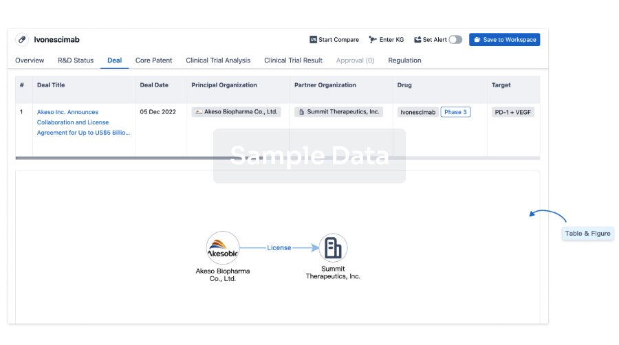
100 Deals associated with COTI-2
Login to view more data
R&D Status
10 top R&D records. to view more data
Login
| Indication | Highest Phase | Country/Location | Organization | Date |
|---|---|---|---|---|
| Colorectal Cancer | Phase 1 | United States | 01 Dec 2015 | |
| Endometrial Carcinoma | Phase 1 | United States | 01 Dec 2015 | |
| Fallopian Tube Carcinoma | Phase 1 | United States | 01 Dec 2015 | |
| Lung Cancer | Phase 1 | United States | 01 Dec 2015 | |
| Ovarian Cancer | Phase 1 | United States | 01 Dec 2015 | |
| Pancreatic Cancer | Phase 1 | United States | 01 Dec 2015 | |
| Peritoneal Neoplasms | Phase 1 | United States | 01 Dec 2015 | |
| Squamous Cell Carcinoma of Head and Neck | Phase 1 | United States | 01 Dec 2015 | |
| Uterine Cervical Cancer | Phase 1 | United States | 01 Dec 2015 | |
| Acute Myeloid Leukemia | Preclinical | United States | - | - |
Login to view more data
Clinical Result
Clinical Result
Indication
Phase
Evaluation
View All Results
| Study | Phase | Population | Analyzed Enrollment | Group | Results | Evaluation | Publication Date |
|---|
No Data | |||||||
Login to view more data
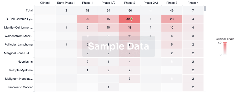
Translational Medicine
Boost your research with our translational medicine data.
login
or

Deal
Boost your decision using our deal data.
login
or

Core Patent
Boost your research with our Core Patent data.
login
or

Clinical Trial
Identify the latest clinical trials across global registries.
login
or

Approval
Accelerate your research with the latest regulatory approval information.
login
or

Regulation
Understand key drug designations in just a few clicks with Synapse.
login
or

AI Agents Built for Biopharma Breakthroughs
Accelerate discovery. Empower decisions. Transform outcomes.
Get started for free today!
Accelerate Strategic R&D decision making with Synapse, PatSnap’s AI-powered Connected Innovation Intelligence Platform Built for Life Sciences Professionals.
Start your data trial now!
Synapse data is also accessible to external entities via APIs or data packages. Empower better decisions with the latest in pharmaceutical intelligence.
Bio
Bio Sequences Search & Analysis
Sign up for free
Chemical
Chemical Structures Search & Analysis
Sign up for free