Request Demo
Last update 29 Mar 2025
NM-01
Last update 29 Mar 2025
Overview
Basic Info
Originator Organization |
Active Organization |
Inactive Organization- |
License Organization- |
Drug Highest PhasePhase 2 |
First Approval Date- |
Regulation- |
Login to view timeline
Related
100 Clinical Results associated with NM-01
Login to view more data
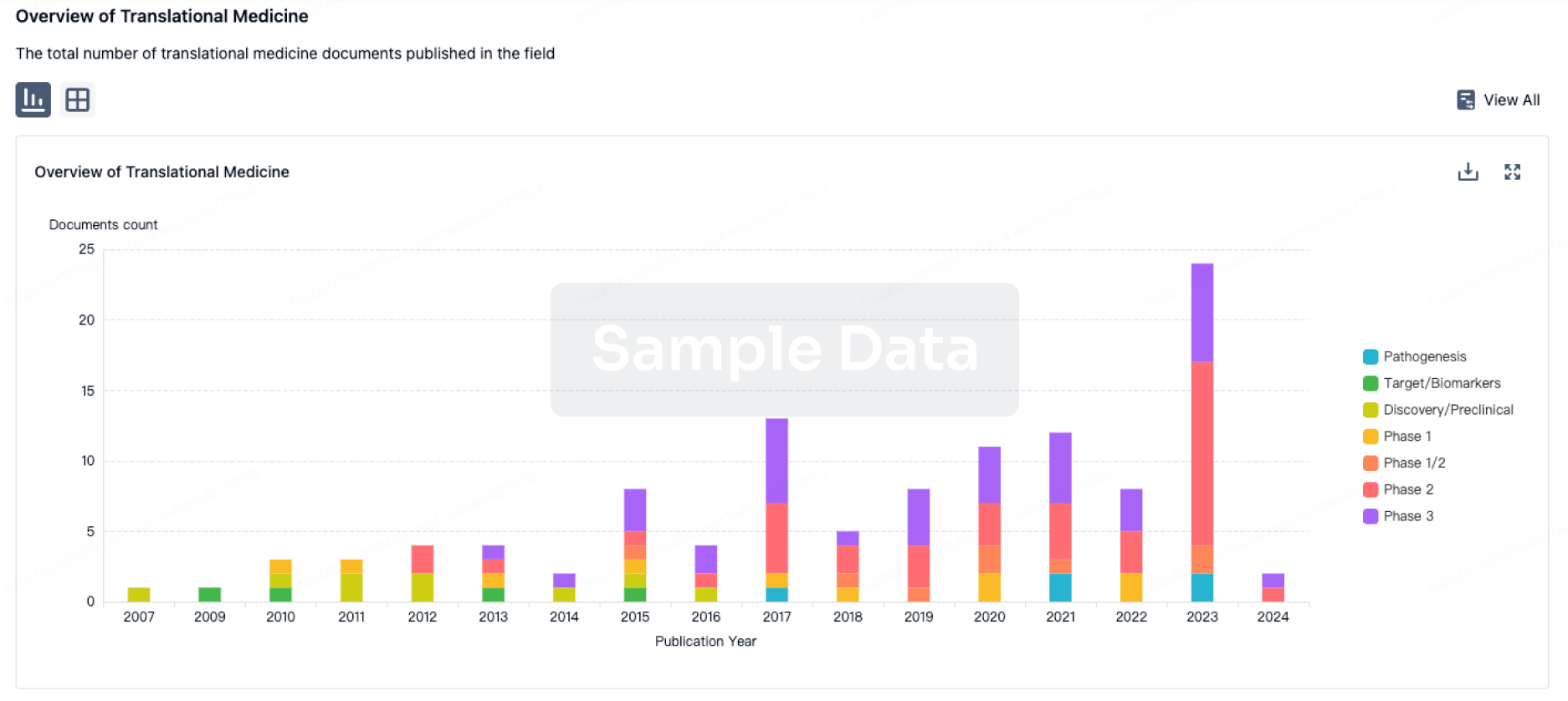
100 Translational Medicine associated with NM-01
Login to view more data
100 Patents (Medical) associated with NM-01
Login to view more data
8
Literatures (Medical) associated with NM-0101 Jan 2021·American journal of nuclear medicine and molecular imaging
Preclinical development and characterisation of 99mTc-NM-01 for SPECT/CT imaging of human PD-L1.
Article
Author: Wong, Nicholas Cl ; Ting, Hong Hoi ; Cai, Yina ; Mottaghy, Felix M ; Meszaros, Levente K ; Cook, Gary Jr ; Biersack, Hans-Jürgen
The level of expression of programmed cell death-1 (PD-1)/programmed death ligand-1 (PD-L1) is a predictive biomarker for cancer immunotherapy, however, its detection remains challenging due to tumour heterogeneity and the influence from the binding of therapeutic agents. We recently developed [99mTc]-NM-01 as a companion diagnostic imaging agent for non-invasive molecular imaging of PD-L1 by single-photon emission computed tomography (SPECT). The aim of the study was to evaluate the [99mTc] radiolabelling of GMP graded NM-01 and its pharmacology, pharmacokinetics and toxicology. NM-01 bound specifically to human PD-L1 (Kd=0.8 nM) and did not interfere with the binding of the anti-PD-L1 antibody atezolizumab. NM-01 can bind various PD-L1-positive cancer cell lines and only interact with PD-L1 expressed on the cell surface. In SPECT/CT imaging, high [99mTc]-NM-01 accumulation was observed in the HCC827 mouse xenografted tumour model (30-min: 1.50 ± 0.27 %ID/g; 90-min: 1.23 ± 0.18 %ID/g), demonstrated a predominantly renal elimination (high uptake in bladder and kidney), while activity in the blood pool and other major organs remained low. The tumour-to-muscle and tumour-to-blood ratios were comparable with/without atezolizumab (P<0.04) but were significantly lowered when co-injected with excess NM-01 (P=0.04 and P=0.01, respectively.) The blood clearance of [99mTc]-NM-01 is bi-phasic; consisting of an initial fast washout phase with half-life of 2.1 min and a slower clearance phase with half-life of 25.4 min. In an intravenous extended single-dose toxicity study, no treatment-related changes were observed and the maximum tolerated dose of [99mTc]-NM-01 was 2.58 mg/kg. [99mTc]-NM-01 has suitable properties as a potential candidate for SPECT/CT imaging of PD-L1 assessment in cancer patients.
01 Sep 2019·Journal of nuclear medicine : official publication, Society of Nuclear MedicineQ1 · MEDICINE
Early Phase I Study of a 99mTc-Labeled Anti–Programmed Death Ligand-1 (PD-L1) Single-Domain Antibody in SPECT/CT Assessment of PD-L1 Expression in Non–Small Cell Lung Cancer
Q1 · MEDICINE
Article
Author: Zhao, Jinhua ; Liu, Changchun ; Ting, Hong Hoi ; O'Doherty, Jim ; Zhao, Lingzhou ; Wong, Nicholas C L ; Meszaros, Levente K ; Xing, Yan ; Cook, Gary J R ; Chand, Gitasha
Immunotherapy with checkpoint inhibitor programmed cell death 1 (PD-1)/programmed death ligand-1 (PD-L1) antibodies demonstrates improvements in treatment of advanced non-small cell lung cancer. Treatment stratification depends on immunohistochemical PD-L1 measurement of biopsy material, an invasive method that does not account for spatiotemporal heterogeneity. Using a single-domain antibody, NM-01, against PD-L1, radiolabeled site-specifically with 99mTc for SPECT imaging, we aimed to assess the safety, radiation dosimetry, and imaging characteristics of this radiopharmaceutical and correlate tumor uptake with PD-L1 immunohistochemistry results. Methods: Sixteen patients (mean age, 61.7 y; 11 men) with non-small cell lung cancer were recruited. Primary tumor PD-L1 expression was measured by immunohistochemistry. NM-01 was radiolabeled with [99mTc(OH2)3(CO)3]+ complex binding to its C-terminal hexahistidine tag. Administered activity was 3.8-10.4 MBq/kg, corresponding to 100 μg or 400 μg of NM-01. Whole-body planar and thoracic SPECT/CT scans were obtained at 1 and 2 h after injection in all patients, and 5 patients underwent additional imaging at 10 min, 3 h, and 24 h for radiation dosimetry calculations. All patients were monitored for adverse events. Results: No drug-related adverse events occurred in this study. The mean effective dose was 8.84 × 10⁻³ ± 9.33 × 10⁻⁴ mSv/MBq (3.59 ± 0.74 mSv per patient). Tracer uptake was observed in the kidneys, spleen, liver, and bone marrow. SPECT primary tumor-to-blood-pool ratios (T:BP) varied from 1.24 to 2.3 (mean, 1.79) at 1 h and 1.24 to 3.53 (mean, 2.22) at 2 h (P = 0.005). Two-hour primary T:BP ratios correlated with PD-L1 immunohistochemistry results (r = 0.68, P = 0.014). Two-hour T:BP was lower in tumors with ≤1% PD-L1 expression (1.89 vs. 2.49, P = 0.048). Nodal and bone metastases showed tracer uptake. Heterogeneity (>20%) between primary tumor and nodal T:BP was present in 4 of 13 patients. Conclusion: This first-in-human study demonstrates that 99mTc-labeled anti-PD-L1-single-domain antibody SPECT/CT imaging is safe and associated with acceptable dosimetry. Tumor uptake is readily visible against background tissues, particularly at 2 h when the T:BP ratio correlates with PD-L1 immunohistochemistry results.
01 Feb 2001·Hybridoma
NM-01 Anti-HIV
Article
2
News (Medical) associated with NM-0104 Aug 2022
Radiopharm will shortly initiate a PD-L1 Phase 1 therapeutic study in Australia in patients with NSCLC
Radiopharm acquired from NanoMab, Ltd. worldwide rights to PD-L1 technology for therapeutic use, as well as to imaging rights in China
MELBOURNE, Australia, Aug. 3, 2022 /PRNewswire/ -- Radiopharm Theranostics (ASX:RAD, "Radiopharm" or the "Company"), a developer of a world-class platform of radiopharmaceutical products for both diagnostic and therapeutic uses, is pleased to announce that it has entered a collaboration agreement with Lantheus for the mutually beneficial development of NM-01, a nanobody made using genetically engineered camelid derived single domain antibodies, that can be labelled with radioisotopes to potentially diagnose and treat multiple tumor types.
In a separate, concurrent agreement, Radiopharm acquired from NanoMab the imaging rights of NM-01 for the strategic Chinese market and worldwide IP rights for any therapeutic use (previously a licencing right).
Radiopharm will shortly initiate a Phase 1 therapeutic trial in Australia in patients with PD-L1 + non-small cell lung cancer (NSCLC). Radiopharm and Lantheus have agreed to cross-reference each other's data to accelerate the development plans for the PD-L1 assets, including the development and regulatory process with USA Food and Drug Administration (FDA) and other key regulatory agencies.
Lantheus provides innovative diagnostics, targeted therapeutics and artificial intelligence (AI) solutions that empower clinicians to Find, Fight and Follow disease. Lantheus holds the exclusive imaging rights to NM-01, apart from China, and recently commenced a Phase 2 clinical trial of NM-01 to evaluate PD-L1 expression in NSCLC patients.
Pursuant to the collaboration agreement, Lantheus will provide the diagnostic product candidate of NM-01 to Radiopharm for use in its therapeutic clinical trials. NM-01 will be used to assess PD-L1 expression during patient selection. In addition, under the agreement, Radiopharm and Lantheus have the option to expand their collaboration to additional assets and potential licensing opportunities in Radiopharm's pipeline.
Radiopharm's CEO & Managing Director Riccardo Canevari said:
"We are excited to have entered a strategically important relationship with Lantheus. We look forward to seeing the results of the Phase 2 PD-L1 imaging trial and to continuing our relationship with Lantheus into the future."
Lantheus' Chief Business Officer, Etienne Montagut said:
"We are pleased to enter into a strategic collaboration with Radiopharm to further the development of NM-01, our novel targeted PD-L1 imaging agent, as a clinical research tool. We believe NM-01's unique potential to evaluate patients before, during, or after treatment with checkpoint inhibitors, will assist Radiopharm in the optimization of the development of its immuno-oncology therapy."
As part of a broader collaboration with NanoMab Ltd, Radiopharm's acquisition of the NanoMab PD-L1 IP will be at no cost for Radiopharm Theranostics.
About the collaboration agreement
Whilst the anticipated expenditure under the collaboration agreement is not considered financially material to Radiopharm in the context of its annual budgeted expenditure, the nature of the agreement and benefit to Radiopharm is considered important, in particular due to Radiopharm:
1) acquiring the imaging rights of NM-01 in the strategic Chinese market
2) acquiring worldwide IP rights of NM-01 for any therapeutic use (previously a licencing right)
3) now has access to data on NM-01 generated by Lantheus to cross reference that data to accelerate the development plans for the PD-L1 assets, including the development and regulatory process with USA Food and Drug Administration (FDA) and other key regulatory agencies.
Expenditure under the agreement is expected to be funded from existing cash reserves. There are no conditions precedent, and the agreement is effective immediately for a term of seven years. The agreement is subject to usual industry termination provisions. Radiopharm has a right of access to information generated during the agreement.
Authorised on behalf of the Radiopharm Theranostics board of directors by Chairman Paul Hopper.
Follow Radiopharm Theranostics:
Website –
Twitter –
Linked In –
SOURCE Radiopharm Theranostics
CollaborateSmall molecular drugAntibodyAcquisition
04 Aug 2022
Radiopharm will shortly initiate a PD-L1 Phase 1 therapeutic study in Australia in patients with NSCLC
Radiopharm acquired from NanoMab, Ltd. worldwide rights to PD-L1 technology for therapeutic use, as well as to imaging rights in China
MELBOURNE, Australia, Aug. 3, 2022 /PRNewswire/ -- Radiopharm Theranostics (ASX:RAD, "Radiopharm" or the "Company"), a developer of a world-class platform of radiopharmaceutical products for both diagnostic and therapeutic uses, is pleased to announce that it has entered a collaboration agreement with Lantheus for the mutually beneficial development of NM-01, a nanobody made using genetically engineered camelid derived single domain antibodies, that can be labelled with radioisotopes to potentially diagnose and treat multiple tumor types.
In a separate, concurrent agreement, Radiopharm acquired from NanoMab the imaging rights of NM-01 for the strategic Chinese market and worldwide IP rights for any therapeutic use (previously a licencing right).
Radiopharm will shortly initiate a Phase 1 therapeutic trial in Australia in patients with PD-L1 + non-small cell lung cancer (NSCLC). Radiopharm and Lantheus have agreed to cross-reference each other's data to accelerate the development plans for the PD-L1 assets, including the development and regulatory process with USA Food and Drug Administration (FDA) and other key regulatory agencies.
Lantheus provides innovative diagnostics, targeted therapeutics and artificial intelligence (AI) solutions that empower clinicians to Find, Fight and Follow disease. Lantheus holds the exclusive imaging rights to NM-01, apart from China, and recently commenced a Phase 2 clinical trial of NM-01 to evaluate PD-L1 expression in NSCLC patients.
Pursuant to the collaboration agreement, Lantheus will provide the diagnostic product candidate of NM-01 to Radiopharm for use in its therapeutic clinical trials. NM-01 will be used to assess PD-L1 expression during patient selection. In addition, under the agreement, Radiopharm and Lantheus have the option to expand their collaboration to additional assets and potential licensing opportunities in Radiopharm's pipeline.
Radiopharm's CEO & Managing Director Riccardo Canevari said:
"We are excited to have entered a strategically important relationship with Lantheus. We look forward to seeing the results of the Phase 2 PD-L1 imaging trial and to continuing our relationship with Lantheus into the future."
Lantheus' Chief Business Officer, Etienne Montagut said:
"We are pleased to enter into a strategic collaboration with Radiopharm to further the development of NM-01, our novel targeted PD-L1 imaging agent, as a clinical research tool. We believe NM-01's unique potential to evaluate patients before, during, or after treatment with checkpoint inhibitors, will assist Radiopharm in the optimization of the development of its immuno-oncology therapy."
As part of a broader collaboration with NanoMab Ltd, Radiopharm's acquisition of the NanoMab PD-L1 IP will be at no cost for Radiopharm Theranostics.
About the collaboration agreement
Whilst the anticipated expenditure under the collaboration agreement is not considered financially material to Radiopharm in the context of its annual budgeted expenditure, the nature of the agreement and benefit to Radiopharm is considered important, in particular due to Radiopharm:
1) acquiring the imaging rights of NM-01 in the strategic Chinese market
2) acquiring worldwide IP rights of NM-01 for any therapeutic use (previously a licencing right)
3) now has access to data on NM-01 generated by Lantheus to cross reference that data to accelerate the development plans for the PD-L1 assets, including the development and regulatory process with USA Food and Drug Administration (FDA) and other key regulatory agencies.
Expenditure under the agreement is expected to be funded from existing cash reserves. There are no conditions precedent, and the agreement is effective immediately for a term of seven years. The agreement is subject to usual industry termination provisions. Radiopharm has a right of access to information generated during the agreement.
Authorised on behalf of the Radiopharm Theranostics board of directors by Chairman Paul Hopper.
Follow Radiopharm Theranostics:
Website –
Twitter –
Linked In –
View original content to download multimedia:
SOURCE Radiopharm Theranostics
Company Codes: Australia:RAD
CollaborateSmall molecular drugAntibodyAcquisition
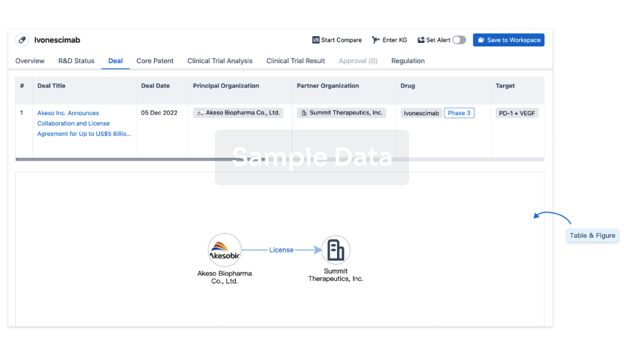
100 Deals associated with NM-01
Login to view more data
R&D Status
10 top R&D records. to view more data
Login
| Indication | Highest Phase | Country/Location | Organization | Date |
|---|---|---|---|---|
| Neoplasms | Phase 2 | United States | 30 Nov 2022 |
Login to view more data
Clinical Result
Clinical Result
Indication
Phase
Evaluation
View All Results
| Study | Phase | Population | Analyzed Enrollment | Group | Results | Evaluation | Publication Date |
|---|
No Data | |||||||
Login to view more data
Translational Medicine
Boost your research with our translational medicine data.
login
or

Deal
Boost your decision using our deal data.
login
or

Core Patent
Boost your research with our Core Patent data.
login
or

Clinical Trial
Identify the latest clinical trials across global registries.
login
or

Approval
Accelerate your research with the latest regulatory approval information.
login
or

Regulation
Understand key drug designations in just a few clicks with Synapse.
login
or

AI Agents Built for Biopharma Breakthroughs
Accelerate discovery. Empower decisions. Transform outcomes.
Get started for free today!
Accelerate Strategic R&D decision making with Synapse, PatSnap’s AI-powered Connected Innovation Intelligence Platform Built for Life Sciences Professionals.
Start your data trial now!
Synapse data is also accessible to external entities via APIs or data packages. Empower better decisions with the latest in pharmaceutical intelligence.
Bio
Bio Sequences Search & Analysis
Sign up for free
Chemical
Chemical Structures Search & Analysis
Sign up for free
