Request Demo
Last update 08 May 2025

Earli, Inc.
Last update 08 May 2025
Overview
Related
100 Clinical Results associated with Earli, Inc.
Login to view more data
0 Patents (Medical) associated with Earli, Inc.
Login to view more data
6
Literatures (Medical) associated with Earli, Inc.21 Apr 2025·Cancer Research
Abstract 60: Nanoparticle delivery of cancer-activated DNA constructs for the diagnosis of liver tumors
Author: Dang, Dang ; Suhy, David ; Wu, Xiaobin ; McCarthy, Blaine ; Chandra, Robby ; Lee, Anderson ; Peterson, Christine ; Lathwal, Sushil ; Cain, Blair ; Morisot, Nadege ; Ananthanarayanan, Badriprasad ; Ramachandran, Suthara ; Wodziak, Dariusz
22 Mar 2024·Cancer Research
Abstract 3529: Leveraging deep learning for fully automated analysis of pre-clinical mouse positron emission tomography
Author: Lee, Hung-Yu Henry ; Sproul, Tim ; Suhy, David ; Goryawala, Mohammed ; Louie, Maggie
22 Mar 2024·Cancer Research
Abstract 4151: Development of a cancer-activated biologic imaging platform for early lung cancer diagnosis
Author: Wodziak, Dariusz ; Xia, Chloe ; Suhy, David ; Sproul, Tim ; Felsher, Dean ; Tong, Ling ; Patil, Trupti ; Lee, Hung-Yu Henry ; Harwig, Alex ; Goryawala, Mohammed ; Ramachandran, Suthara
4
News (Medical) associated with Earli, Inc.21 Aug 2024
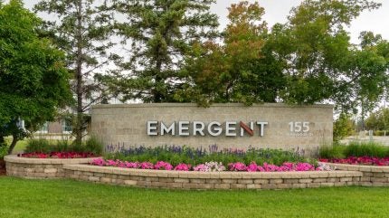
Emergent’s facility sale comes as the company attempts to salvage its teetering financial situation. Image: Shutterstock/ JHVEPhoto.
Emergent BioSolutions
announced that it has finalised the sale of its Baltimore-Camden manufacturing site to the Taiwanese firm Bora Pharmaceuticals.
The sale was first
announced in July 2024
, as Emergent sought to recover from three years of financial decline following initial commercial success during the Covid-19 pandemic. Emergent is set to receive approximately $30m from Bora, subject to post-closing adjustments. In return, Bora will acquire the assets, equipment, and workforce of the 87,000 square foot facility.
Joe Papa, President and CEO of Emergent described the sale as, “a significant step forward in our multi-year plan to stabilise, turnaround, and transform Emergent,” as the company moves towards becoming a “leaner and more flexible organisation”. Papa stated that the development contributes to a “multi-year plan to improve overall profitability and raise capital to reduce our debt.”
According to
Yahoo Finance
, Emergent share price had declined by as much as 98.85% by February 2024 from a high during August 2020. However, more recently, after the World Health Organization declared a global public health emergency of international concern regarding mpox, the company’s stock has risen by 41.83%. Emergent’s market cap currently stands at $514m.
On 19 August, the company announced it would donate 50,000 doses of their ACAM2000 vaccine to the Democratic Republic of Congo and other nations affected by mpox outbreaks. The vaccine,
acquired from Sanofi
in 2017, can be used for mpox immunisation under an Expanded Access Investigational New Drug (EA-IND) protocol.
See Also:
Innovent’s Dupert approved in China to treat lung cancer
Accenture invests in Earli to advance early cancer detection tech
GlobalData projects ACAM2000 will generate $121m in revenue in 2024, increasing to $129m annually by 2030.
GlobalData is the parent company of
Pharmaceutical Technology.
The Bora deal follows Emergent’s sale of its RSDL (Reactive Skin Decontaminant Lotion) kit to SERB Pharmaceuticals in July 2024 for $75m. The company also announced in May that it would eliminate 300 jobs and shut down two facilities in Baltimore and Maryland to save an estimated $80m in annual costs. This year, Emergent has also agreed on deals with the US government worth as much as $488m to supply vaccines and other therapeutics. In the meantime, the company maintains focus on its blockbuster opioid overdose medication Narcan (naloxone).
Emergent’s sale represents the second US facility acquisition by Bora, following its purchase of Upsher-Smith Laboratories in April this year. The Taiwanese firm is expanding its contract development and manufacturing and organisation (CDMO) operations into the US as the BIOSECURE Act threatens American corporate ties to
many of the company’s rivals, particularly in China
.

VaccineAcquisitionDrug Approval
20 Aug 2024
The synthetic biopsy method is designed to improve the sensitivity and specificity of cancer detection. Credit: Chinnapong/Shutterstock.
Accenture
has invested in biotechnology company Earli to advance early cancer detection technologies.
Facilitated through Accenture Ventures, the investment aims to enhance collaborations with global health and pharma companies, leveraging Earli’s synthetic targeting platform that reprogrammes cancer cells to aid in their detection and destruction.
Earli’s technology focuses on the early and accurate distinction between healthy and cancerous cells.
The synthetic biopsy method is designed to improve the sensitivity and specificity of cancer detection, potentially leading to earlier diagnoses and more personalised treatment options.
Earli’s technology is poised to revolutionise cancer detection by using non-invasive screening methods, such as blood samples and PET [positron emission topography] scans, to identify multiple cancer types.
See Also:
FDA approves updated mRNA vaccines for Covid-19
Verve Therapeutics gets grant for gene editing compositions to lower ldl cholesterol levels
The investment from Accenture Ventures is expected to help Earli expand its collaborations, potentially accelerating the development and availability of early cancer detection and treatment solutions.
Earli joins Accenture Ventures’ engagement and investment programme, Project Spotlight, working with startups on disruptive enterprise technologies.
Accenture Life Sciences business senior managing director and global lead Petra Jantzer stated: “Earli’s synthetic biopsy method is a step change in early cancer detection technologies and will offer significant advantages to biopharma companies by improving the precision and efficacy of new treatments and diagnostics, as well as in understanding the mechanisms of cancer progressions.”
Project Spotlight provides access to the domain expertise of Accenture as well as its enterprise clients.
Biotechnology companies Turbine, QuantHealth, Virtonomy and Ocean Genomics have also joined Project Spotlight.
Accenture Ventures global lead Tom Lounibos said: “With Earli joining our Project Spotlight programme, we can collaborate with our clients in the biopharma industry to advance their capabilities in cancer research, drug development and patient care.”
VaccinemRNADrug Approval
15 Jan 2021
Although the 39th annual J.P. Morgan Healthcare Conference looked quite a bit different than the 38 before it held in San Francisco, the virtual conference still held plenty of wheeling and dealing for the biotech world. Here are the companies who found their zoom meetings with investors quite profitable.
Tessera Therapeutics
Unveiled in July after years of working in stealth mode, Tessera is rewriting our genes to treat disease. Authoring therapeutic instructions into the genome takes cash and lots of it. This week Tessera brought in over $230 million in a Series B. By changing any gene base pair to another, inserting or deleting, and writing entire genes into the genome, the company hopes to unlock potential to cure genetic diseases and create life-changing therapeutics in cardiovascular, oncological, neurodegenerative and infectious diseases. The funds will be used to accelerate research and development in the company’s gene writing technologies, expand its team and establish manufacturing and automation capabilities critical for its platform and programs.
EQRx
This Cambridge startup is looking to turn the drug industry on its head by bringing new, life-saving medicines to patients at a fraction of the cost of today’s leading therapies. The “remaking medicine” company announced a $500 million Series B financing to further their worthy cause. EQRx is busy building a highly competitive pipeline of drugs that has the potential to save the US healthcare system between 50-70% of its current drug spend. Initial targets are candidates for cancer and inflammatory diseases. Several late-stage drugs currently in development show promise in some of the most common cancers – lung, breast and other solid tumors.
NewAmsterdam
Netherlands’ NewAmsterdam Pharma hauled in a healthy Series A with $196 million in funding. The clinical stage company is focused on therapy development for cardio-metabolic diseases. This chunk of change will help the company take its small molecule drug, obicetrapib, into full Phase III development. The drug is a cholesteryl ester transfer protein inhibitor for patients not well-controlled on statins. NewAmsterdam’s founding investor Forbion participated along with Peter Thiel, PayPal co-founder and billionaire venture capitalist.
Sana Biotechnology
Gene editor Sana is dipping its toe into the Nasdaq, filing Wednesday to go public with the goal of raising up to $150 million. “Our long-term aspirations are to be able to control or modify any gene in the body, to replace any cell that is damaged or missing, and to markedly improve access to cellular and gene-based medicines,” the company wrote in its IPO prospectus. The approach has potential with a number of disease,s but most of the advanced research at Sana right now is focused on cancer. The company anticipated filing NDAs for multiple therapies soon, starting as early as 2022.
Delfi Diagnostics
Potentially lowering the cost of cancer detection while identifying it earlier is the goal of Delfi’s tech, based off the research of founder and CEO Victor Velculescu. Delfi brought in a $100 million Series A round. Enhanced by machine learning technology, the biotech is developing cell-free DNA-based liquid biopsies to detect the presence of tumors and the tumor’s tissue of origin. The Series A funds will be used to build Delfi’s team and launch validation studies for its tech.
Valo Health
Working to transform drug discovery and development process, Valo unveiled select therapeutics programs and closed a $190 million Series B financing round. Combining the power of patient data and machine learning technology has allowed the rapid development of key preclinical programs. The company’s oncology portfolio includes hematological and solid tumor malignancies, brain tumors, c-myc driven cancers and particular solid tumors.
Atalanta Therapeutics
Boston-based Atalanta launched this week with $110 million in combined Series A funding and collaboration deals with Genentech and Biogen to address diseases related to the central nervous system, including Huntington’s, Alzheimer’s and Parkinson’s diseases. The company believes its approach developing RNAi drugs using branched siRNA, a new type of molecular architecture, has the potential to overcome the challenges of brain and spinal cord medicine distribution. Preclinical research has shown that branched siRNA can achieve what Atalanta called “unparalleled distribution in the CNS,” which includes deep brain structures and prolonged duration of effect.
Elucida Oncology
Drug conjugate company Elucida is targeting ovarian and brain cancers. The company’s C-Dots can precisely target and penetrate tumors to deliver the drug payload, then be safely cleared by the kidneys. With an additional $44 million in a Series A-1, the Elucida’s Series A total is now $72 million. The funding will be used to complete IND studies for the company’s lead candidate to get it into the clinic by late 2021, according to the CEO. Elucida has also partnered up to develop diagnostics and surgical applications based on the technology.
IO Biotech
Granted breakthrough therapy designation by the FDA just last month, IO Biotech hit it big with over a $154 million Series B financing round. The designation is for the company’s combo therapies IO102 and IO103 with anti-PD-1 monoclonal antibodies for patients with unresectable or metastatic melanoma. The designation will help expedite the development and review of IO’s drugs. The biotech intends to use the net proceeds of the transaction towards the funding of clinical trials for its early and late-stage immuno-oncology programs, including a large randomized trial for IO102 and IO103 with anti-PD-1 monoclonal antibodies in metastatic melanoma.
DiCE Molecules
In a Series C financing round, DiCE secured $80 million to support the development of its first-in-class, oral IL-17 agonist and roll it into the clinic. The IL-17 family of cytokines are strong inducers of inflammation and are implicated in a variety of autoimmune diseases including psoriasis, psoriatic arthritis and ankylosing spondylitis. Other assets benefiting from the fund include a pair of integrin inhibitors, and plans to expand the pipeline using the same combo of technology and structural insights. “We believe the immunology space is underserved by current small molecule approaches and we are excited about the opportunity to advance next-generation therapeutics for this patient population,” Judice said in a statement.
Earli
This bioengineering firm developed a new platform technology to give cancer patients their best chance at survival – early detection. With tech licensed from Standford, Earli is creating a platform called “Synthetic Biopsy.” The platform uses genetic constructs to force cancer to produce biomarkers not normally expressed in the human body. Clinicians can then exactly locate early cancers to begin treatment. After a $19.5 million seed investment in 2018, the biotech has now raised $40 million in a Series A round. "The Earli platform is radically different from other early cancer detection and treatment approaches. It makes early detection localizable and therefore actionable, which is the critical next step needed for success against cancer," said Vinod Khosla, founder of Khosla Ventures who led the financing round. “We believe Earli has the potential to forever change the cancer playbook.”
AntibodyBreakthrough TherapyFirst in ClassSmall molecular drugCollaborate
100 Deals associated with Earli, Inc.
Login to view more data
100 Translational Medicine associated with Earli, Inc.
Login to view more data
Corporation Tree
Boost your research with our corporation tree data.
login
or

Pipeline
Pipeline Snapshot as of 07 Feb 2026
No data posted
Login to keep update
Deal
Boost your decision using our deal data.
login
or

Translational Medicine
Boost your research with our translational medicine data.
login
or

Profit
Explore the financial positions of over 360K organizations with Synapse.
login
or

Grant & Funding(NIH)
Access more than 2 million grant and funding information to elevate your research journey.
login
or

Investment
Gain insights on the latest company investments from start-ups to established corporations.
login
or

Financing
Unearth financing trends to validate and advance investment opportunities.
login
or

AI Agents Built for Biopharma Breakthroughs
Accelerate discovery. Empower decisions. Transform outcomes.
Get started for free today!
Accelerate Strategic R&D decision making with Synapse, PatSnap’s AI-powered Connected Innovation Intelligence Platform Built for Life Sciences Professionals.
Start your data trial now!
Synapse data is also accessible to external entities via APIs or data packages. Empower better decisions with the latest in pharmaceutical intelligence.
Bio
Bio Sequences Search & Analysis
Sign up for free
Chemical
Chemical Structures Search & Analysis
Sign up for free