Request Demo
Last update 08 May 2025

New York Methodist Hospital
Last update 08 May 2025
Overview
Related
100 Clinical Results associated with New York Methodist Hospital
Login to view more data
0 Patents (Medical) associated with New York Methodist Hospital
Login to view more data
1,158
Literatures (Medical) associated with New York Methodist Hospital12 Nov 2024·Circulation
Abstract 4139444: Benign Metastasizing Leiomyoma: A Challenging Case with Multifocal Involvement in the Heart, Liver, and Uterus
Author: Vu, Huong ; Minh, Le Huu Nhat ; Vo, Duc ; Huynh, Hai ; Nguyen, Hien ; Bui, Hanh
01 May 2024·Canadian Journal of Emergency Medicine
CJEM Debate Series: #TriageAgain—are current triage methods dangerous?… if we cannot actually treat those triaged as urgent within a safe time frame?
Author: Atkinson, Paul ; Melniker, Larry ; Ballesteros, Patrick ; Davies, Ffion
01 May 2024·A66. BACTERIA CASES UNLEASHED
Management of Complicated Empyema With Urgent Video Assisted Thoracoscopic Surgery (VATS)
Author: Haile, Y. ; Seylani, A. ; Tran, L.-K.
3
News (Medical) associated with New York Methodist Hospital07 Nov 2023
TARRYTOWN, N.Y., Nov. 7, 2023 /PRNewswire/ -- Prominently featured in The Inner Circle, Pierre-Yves Sonke, MD, is acknowledged as a Top Pinnacle Healthcare Professional for his contributions to Medical Imaging.
Continue Reading
Pierre-Yves Sonke
Dr. Sonke began his scholarship at Erasmus Medical School in Rotterdam Netherlands and graduated in 1985. He then continued with a two-year fellowship in cancer research focused in radiation biology supported by the Kankerbestrijding (KWF), Dutch Cancer Society, in Rijswijk and Amsterdam Netherlands. Upon relocating to England, he completed internships in oncology at Princess Royal Hospital; emergency medicine at Hull Royal Infirmary; anesthesia at Royal Berkshire Hospital; and clinical chemistry at Royal Hallamshire Hospital.
Continuing his exceptional education, Dr. Sonke enrolled a residency in anatomical and clinical pathology in Long Island, New York. He completed a residency in nuclear medicine at North Shore Hospital and in diagnostic radiology at New York Methodist Hospital. The doctor attained board certification in diagnostic radiology through ABR and notes that the ABR is a not-for-profit physician-led organization that oversees the certification and ongoing professional development of specialists in diagnostic radiology interventional radiology radiation oncology and medical physics.
Dr. Sonke is currently an attending physician at Westchester Medical Center in Valhalla, New York. He is considered a specialist, including body imaging; nuclear medicine; PET/CT; body CT; MR and ultrasound imaging. According to the doctor, diagnostic radiology refers to the field of medicine that uses non-invasive imaging scans for medical diagnosis.
In addition to his medical practice, Dr. Sonke is an assistant professor of radiology at New York Medical College. He is a member of the American Roentgen Ray Society and the European Society of Radiology.
Contact: Katherine Green, 516-825-5634, editorial@continentalwhoswho.com
SOURCE The Inner Circle
Radiation TherapyExecutive Change
14 Aug 2023
TARRYTOWN, N.Y., Aug. 14, 2023 /PRNewswire/ -- Prominently featured in The Inner Circle, Pierre-Yves Sonke, MD, is acknowledged as a Top Pinnacle Healthcare Professional for his contributions to Medical Imaging.
Continue Reading
Pierre-Yves Sonke
Dr. Sonke began his scholarship at Erasmus Medical School in Rotterdam Netherlands and graduated in 1985. He then continued with a two-year fellowship in cancer research focused in radiation biology supported by the Kankerbestrijding (KWF), Dutch Cancer Society, in Rijswijk and Amsterdam Netherlands. Upon relocating to England, he completed internships in oncology at Princess Royal Hospital; emergency medicine at Hull Royal Infirmary; anesthesia at Royal Berkshire Hospital; and clinical chemistry at Royal Hallamshire Hospital.
Continuing his exceptional education, Dr. Sonke enrolled a residency in anatomical and clinical pathology in Long Island, New York. He completed a residency in nuclear medicine at North Shore Hospital and in diagnostic radiology at New York Methodist Hospital. The doctor attained board certification in diagnostic radiology through ABR and notes that the ABR is a not-for-profit physician-led organization that oversees the certification and ongoing professional development of specialists in diagnostic radiology interventional radiology radiation oncology and medical physics.
Dr. Sonke is currently an attending physician at Westchester Medical Center in Valhalla, New York. He is considered a specialist, including body imaging; nuclear medicine; PET/CT; body CT; MR and ultrasound imaging. According to the doctor, diagnostic radiology refers to the field of medicine that uses non-invasive imaging scans for medical diagnosis.
In addition to his medical practice, Dr. Sonke is an assistant professor of radiology at New York Medical College. He is a member of the American Roentgen Ray Society and the European Society of Radiology.
Contact: Katherine Green, 516-825-5634, [email protected]
SOURCE The Inner Circle
Radiation TherapyExecutive Change
28 Jun 2022
BROOKLYN, N.Y., June 28, 2022 /PRNewswire/ -- Rizwanullah Hameed, MD, is being recognized by Continental Who's Who as a Pinnacle Healthcare Professional for his excellence in the Medical field and in acknowledgment of his private primary care practice.
A board-certified physician specializing in Internal Medicine and Infectious Disease, Dr. Rizwanullah Hameed has provided high-quality medical care to patients in the New York City region for over 30 years. He specializes in preventing, diagnosing, and treating various infectious diseases.
Continue Reading
Rizwanullah Hameed
Alongside his primary care practice, Dr. Hameed is affiliated with Kingsbrook Jewish Medical Center and New York Methodist Hospital. He has served as Chairman of the Hospital Pharmaceutical and Therapeutic Committee, Medical Staff President, Chief of Infectious Diseases, and IRB committee member at Kingsbrook Jewish Medical Center. He has also served as Vice-Chairman of Community Medicine at NY Methodist Hospital.
In addition to his clinical work, Dr. Hameed has published seven clinical and epidemiology studies and has completed numerous clinical research courses.
In pursuit of his medical career, Dr. Hameed studied medicine at Rawalpindi Medical University in Pakistan, where he graduated in 1984. After relocating to the US, he completed an Internal Medicine Residency at Kingsbrook Jewish Medical Center, in Brooklyn, in 1991 and a Fellowship in Infectious Diseases at Harlem Hospital and Columbia University in New York City in 1993.
In accepting this honorable recognition, Dr. Hameed would like to thank his mentor, Dr. Martin Kramer. He would also like to thank his wife, Sameeram, their children, Saad and Marvi, and grandson, Saad, and give loving remembrance to his late parents.
For more information, visit .
Contact: Katherine Green, 516-825-5634, [email protected]
SOURCE Continental Who's Who
100 Deals associated with New York Methodist Hospital
Login to view more data
100 Translational Medicine associated with New York Methodist Hospital
Login to view more data
Corporation Tree
Boost your research with our corporation tree data.
login
or

Pipeline
Pipeline Snapshot as of 15 May 2025
No data posted
Login to keep update
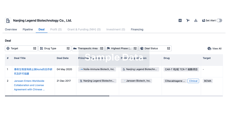
Deal
Boost your decision using our deal data.
login
or
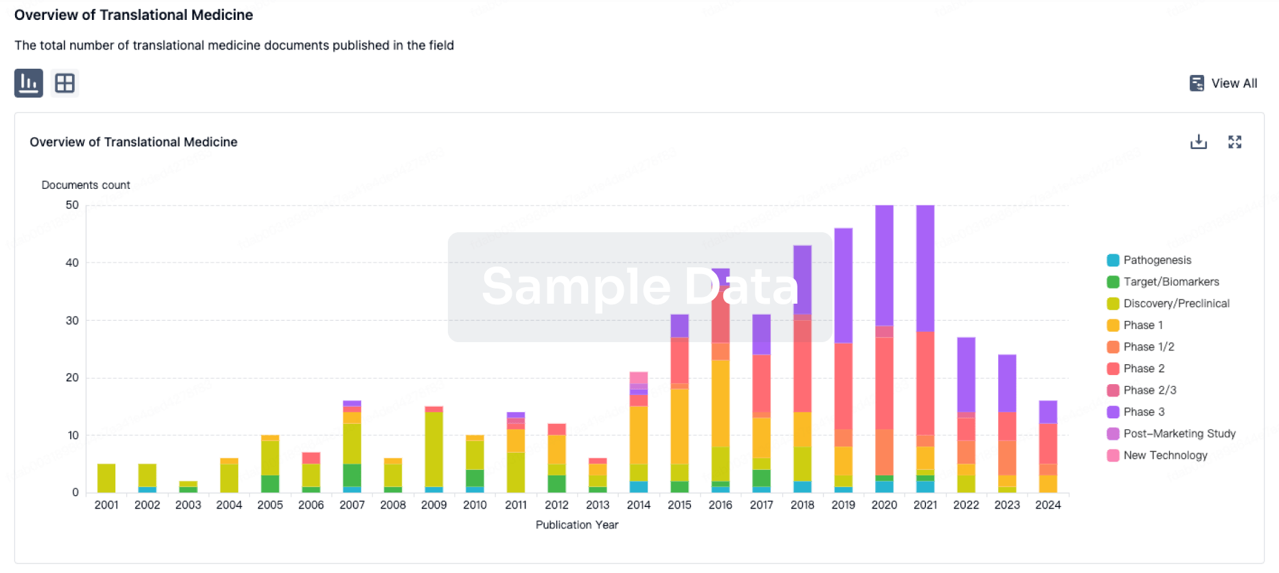
Translational Medicine
Boost your research with our translational medicine data.
login
or
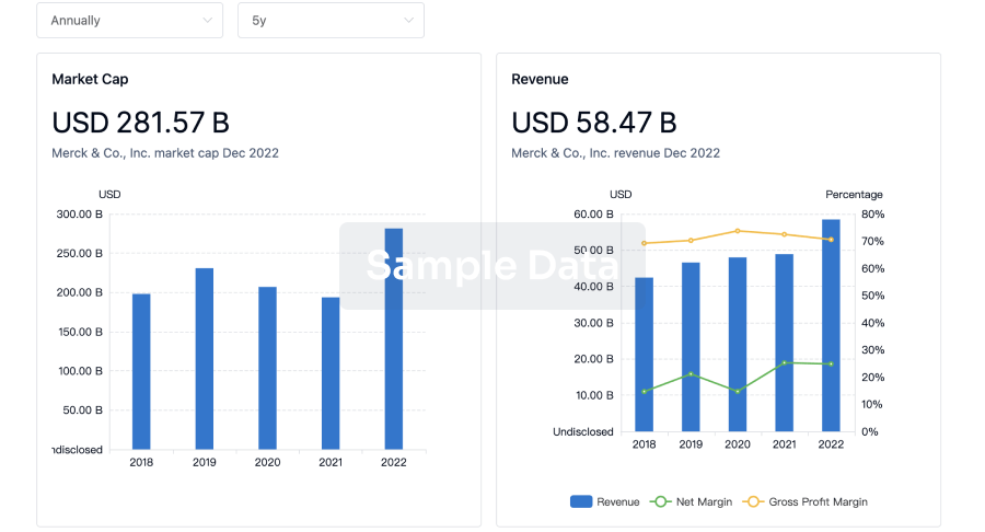
Profit
Explore the financial positions of over 360K organizations with Synapse.
login
or
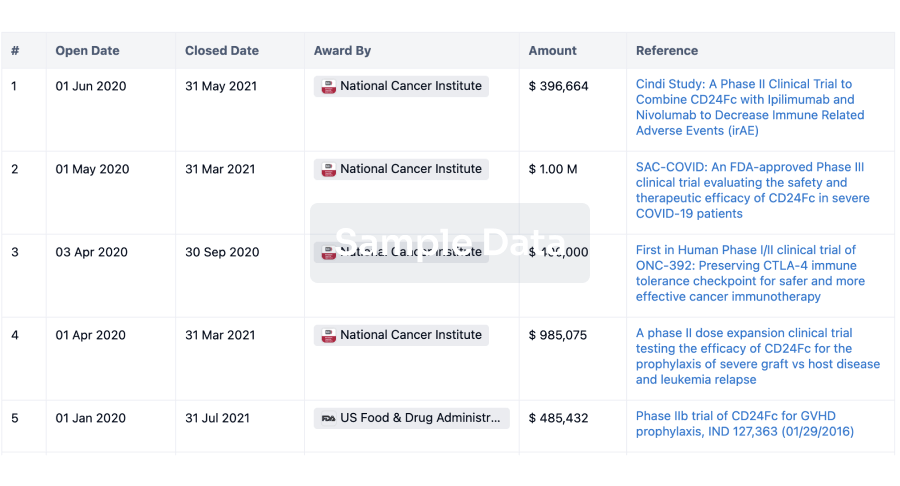
Grant & Funding(NIH)
Access more than 2 million grant and funding information to elevate your research journey.
login
or
Investment
Gain insights on the latest company investments from start-ups to established corporations.
login
or
Financing
Unearth financing trends to validate and advance investment opportunities.
login
or
Chat with Hiro
Get started for free today!
Accelerate Strategic R&D decision making with Synapse, PatSnap’s AI-powered Connected Innovation Intelligence Platform Built for Life Sciences Professionals.
Start your data trial now!
Synapse data is also accessible to external entities via APIs or data packages. Empower better decisions with the latest in pharmaceutical intelligence.
Bio
Bio Sequences Search & Analysis
Sign up for free
Chemical
Chemical Structures Search & Analysis
Sign up for free