Request Demo
Last update 08 May 2025
Vizgen, Inc.
Last update 08 May 2025
Overview
Related
100 Clinical Results associated with Vizgen, Inc.
Login to view more data
0 Patents (Medical) associated with Vizgen, Inc.
Login to view more data
34
Literatures (Medical) associated with Vizgen, Inc.21 Apr 2025·Cancer Research
Abstract 5809: Spatially resolved immunologic hallmarks of response to neoadjuvant immune checkpoint blockade in metastatic melanoma
Author: Moran-Segura, Carlos ; He, Justin ; Johnson, Joseph ; Marchion, Douglas ; Sayegh, Zena ; Nguyen, Jonathan ; Balasi, Jodi ; Chuang, Jeffrey H. ; Zhang, Chaomei ; Liu, Zichao ; Song, Xiaofei ; Messina, Jane ; He, Jiang ; Sondak, Vernon K. ; Yoder, Sean ; Chen, Pei-Ling ; Chen, Wei-Shen ; Sun, Nan
21 Apr 2025·Cancer Research
Abstract 6310: Bridging spatial proteomics and classical histopathology: advanced spatial analysis through H&E-enhanced multiplexed immunoprofiling
Author: Cardenes, Ruben ; Wood, Douglas ; Rognoni, Lorenz ; Downing, Michael ; Chatterjee, Gourab
19 Jul 2024·Clinical and Experimental Dermatology
Deep resolution of clinical, cellular and transcriptomic inflammatory markers of psoriasis over 52 weeks of interleukin-17A inhibition by secukinumab
Article
Author: Krueger, James G ; Suárez-Fariñas, Mayte ; Kolbinger, Frank ; Hartmann, Nicole ; Peters, Thomas ; Milutinovic, Marina ; Glueck, Anton ; Tomalin, Lewis E ; Suprun, Maria ; Wharton, Keith A
43
News (Medical) associated with Vizgen, Inc.09 Oct 2024
Vizgen and Ultivue, two companies at the forefront of developing technologies that allow scientists to map the molecular makeup of cells in exquisite detail, are joining forces.
The merger,
announced
Wednesday, creates the biggest private company in the emerging spatial biology field. The startups have raised more than $256 million total over the years.
That sum doesn’t include a new Series D financing for Vizgen. The company would not disclose the amount raised but said it was backed by ARCH Venture Partners, Northpond Ventures and Tao Capital Partners. Those three firms have also previously invested in Ultivue.
Vizgen’s instruments map which RNA molecules are present in individual cells across a tissue, a technique broadly known as spatial transcriptomics. Ultivue sells kits to detect up to a dozen different proteins on a single tissue sample, which is one version of a method known as spatial proteomics.
Scientists are using the techniques to produce kaleidoscopic images
revealing the complexity of the brain
, the immune system and the tumor microenvironment. It’s leading to new hypotheses about what goes awry in Alzheimer’s disease, autoimmune diseases, cancer and more. Researchers hope the approaches will lead to new drugs and diagnostics. Yet the booming field of spatial biology has been rocked by a series of lawsuits, including one directed at Vizgen.
The California company 10X Genomics has sued Vizgen for infringing its patents. Vizgen has denied the allegation, countersued, and accused 10X of trying to quash competition with monopolistic practices.
In a written statement, Vizgen told
Endpoints News
that the merger “does not relate to our ongoing litigation” and that the company will continue to defend its technology. A trial is scheduled for early 2025.
“Customers can expect continued access to the same products and services with added benefits from the combined expertise and technology platforms of both Vizgen and Ultivue,” the company told Endpoints.
Vizgen and Ultivue are based just blocks apart in the same industrial park on the western outskirts of Cambridge, MA. The companies will maintain their presence there, as well as Vizgen’s manufacturing facility in nearby Waltham, MA. Vizgen said that it hasn’t announced layoffs.
Ultivue CEO Rob Carson will run the combined company, which will operate under the Vizgen name. Vizgen CEO Terry Lo will remain on the company’s board. Carson and Lo declined interview requests through a spokesperson.
Earlier this year, NanoString, another spatial biology company, declared bankruptcy in the midst of its own legal dispute with 10X.
10X, NanoString and Vizgen all sell instruments for spatial transcriptomics. The details of each company’s approach vary, but all of them give researchers a picture of how gene expression varies from cell-to-cell across a tissue.
Some academic scientists who rely on spatial biology instruments from these companies have also accused 10X of trying to stifle innovation — 10X has
denied it
— and voiced concerns that their investments in expensive instruments from NanoString and Vizgen will be useless if the companies fold.
NanoString CEO Brad Grey told Endpoints in April that the company’s debt and legal losses
made it impossible to raise money
to keep the company afloat and cover the $31 million in damages that US courts said it owed 10X. In May, the scientific instrument company Bruker said it would
acquire
NanoString for $392.6 million in cash.
On Wednesday, Bruker
announced
a new division of the company focused on spatial biology that will continue to offer NanoString instruments and reagents alongside contract research services.
Acquisition
08 Oct 2024
CAMBRIDGE, Mass., October 9, 2024 – Vizgen, Inc., a life science company dedicated to improving human health by visualizing single-cell spatial genomics information, and Ultivue, Inc., a market leader in multiplex proteomic biomarker detection and advanced AI-driven spatial tissue profiling, today announced a merger. The companies are combining to establish a single organization uniquely positioned to drive future technology breakthroughs and respond to researchers’ desire for unified solutions in spatial multi-omics.
“This merger represents a pivotal moment in the evolution of spatial multi-omics. By bringing together Vizgen’s expertise in single-cell spatial genomics and Ultivue’s leadership in multiplex proteomic profiling, we are unlocking new opportunities for innovation in discovery and clinical development,” said Andrea Jackson, Partner at Northpond Ventures. “I have tremendous confidence in the combined team’s ability to further build on their technology leadership and increased scale.”
Rob Carson, formerly CEO of Ultivue, will become the President and CEO of the newly-combined Vizgen. Terry Lo, who served as Vizgen’s CEO beginning in 2020 and led the company through a period of innovation, global expansion and significant growth, will continue to serve as a member of the board of directors.
The Vizgen-Ultivue merger brings together two established spatial biology companies, uniting Vizgen’s breakthrough MERFISH spatial genomics technology – including its MERSCOPE Ultra™ Spatial Imaging platform – with Ultivue’s high-fidelity InSituPlex® assays and AI-powered STARVUE™ platform. These proven technologies provide researchers with the sub-cellular precision required to understand interactions at the molecular level.
“Imagine having an exquisitely detailed map that not only shows you where cells are, but also how they are interacting with their environment, what they are expressing, and how they are responding to genetic plans and disease treatments. Spatial multi-omics is like having a crystal-clear window into complex cellular interactions that reveals how gene expression and protein function interact to change human biology,” said Jiang He, PhD, Vice President Reagents & Consumables, Scientific Co-Founder of Vizgen.
Carson, who has more than 20 years of experience in the life sciences, including business unit and enterprise leadership roles at Medtronic and Waters Corporation, will unite talented teams with extensive and complementary expertise in molecular biology and engineering. He and the Ultivue team previously led significant commercial growth, product portfolio expansion and operational improvements.
“I’m very excited to work alongside both existing and new colleagues here at Vizgen as we help to write new chapters on what’s possible through spatial biology. We’re positioned to serve research applications across the spatial spectrum, drawing on our customers’ experiences and insights, our team’s synergistic skill sets, and two high performance platform technologies,” said Carson.
Concurrent with the merger, Vizgen also announced completion of its Series D round of new equity financing. The company continues to be supported by a global syndicate of leading investors in the life sciences, including ARCH Venture Partners, Northpond Ventures and Tao Capital Partners, among others.
Vizgen is at the forefront of spatial biology and multi-omics innovation. Through its pioneering MERFISH technology and MERSCOPE® Platform for in situ single-cell spatial genomics and its high-fidelity InSituPlex® assays and AI-driven STARVUE™ spatial image analysis technology, Vizgen delivers unmatched tools that enable researchers to uncover deep insights into human biology and achieve breakthroughs in understanding mechanisms of complex diseases. Vizgen’s technology delivers high-confidence, quantitative data with exceptional accuracy, robustness, sensitivity and deep insights that power foundational and clinical research. Vizgen is headquartered in Cambridge, Massachusetts, and can be found online at www.vizgen.com or www.ultivue.com.
Executive ChangeAcquisition
16 Apr 2024
CAMBRIDGE, Mass.--(BUSINESS WIRE)-- Vizgen, Inc., the life science company dedicated to improving human health by visualizing single-cell spatial genomics information, announced today the issuance by the US Patent Office of a patent that covers the in situ imaging of the spatial transcriptome. U.S. Patent No. 11,959,075 is for robust multiplex imaging of RNA and its spatial organization in a sample.
"This latest patent relates to our MERFISH technology and enables groundbreaking advancements in the spatial genomics landscape," said Terry Lo, CEO, Vizgen, Inc. "It is another patent underscoring Vizgen’s leadership in the field, providing the industry’s best tools for single-cell spatial biology, and driving discoveries aimed at improving human health," he said.
Vizgen has secured key patents protecting its innovative pioneering technology in the United States and abroad, adding to its growing patent portfolio as Vizgen continues to invest in the development of its groundbreaking technologies. This newly allowed patent is owned by The President and Fellows of Harvard College and is exclusively licensed to Vizgen.
Reflecting its long-term commitment to scientific innovation and biomedical progress, and despite attempts by others to monopolize the field by aggressive litigation tactics, Vizgen is making the newly issued patent available for licensing.
The issuance of the ‘075 Patent comes just over a month after the PTAB denied institution of an Inter Partes Review filed by 10X Genomics, Inc. of the parent patent, U.S. Patent No. 11,098,303 also directed to the pioneering technology invented by Dr. Xiaowei Zhuang at Harvard University.
To date, there are over two hundred peer-reviewed and preprint publications on MERFISH technology and thousands of samples have been processed on MERSCOPE instruments. During AACR 2024, Vizgen unveiled the MERSCOPE® Ultra Platform and shared new MERFISH 2.0 chemistry data. These powerful tools are uniquely suited to supporting basic and translational research and will be commercially available in the second half of the year.
About Vizgen®
Vizgen is dedicated to pioneering the next generation of genomics, providing tools that demonstrate the possibilities of in situ single-cell spatial genomics. The company has established the benchmark in spatial genomics and continues to drive industry innovation, delivering the highest sensitivity as well as excellent specificity and accuracy in cell segmentation. These tools enable researchers to gain new insight into the biological systems that govern human health and disease with spatial context. Vizgen’s MERSCOPE® Platform enables massively multiplexed, genome-scale nucleic acid imaging with high accuracy and unrivaled detection efficiency at subcellular resolution. MERSCOPE provides transformative insight into a wide range of tissue-scale basic research and translational medicine in oncology, immunology, neuroscience, infectious disease, developmental biology, cell and gene therapy, and is an essential tool for accelerating drug discovery and development. For more information, go to . Connect on social media Twitter, LinkedIn and Facebook.
100 Deals associated with Vizgen, Inc.
Login to view more data
100 Translational Medicine associated with Vizgen, Inc.
Login to view more data
Corporation Tree
Boost your research with our corporation tree data.
login
or

Pipeline
Pipeline Snapshot as of 19 Dec 2025
No data posted
Login to keep update
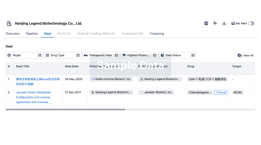
Deal
Boost your decision using our deal data.
login
or
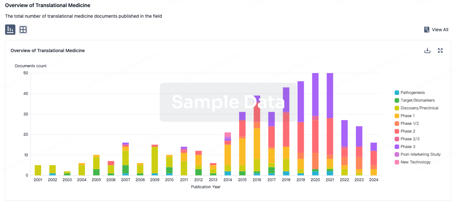
Translational Medicine
Boost your research with our translational medicine data.
login
or
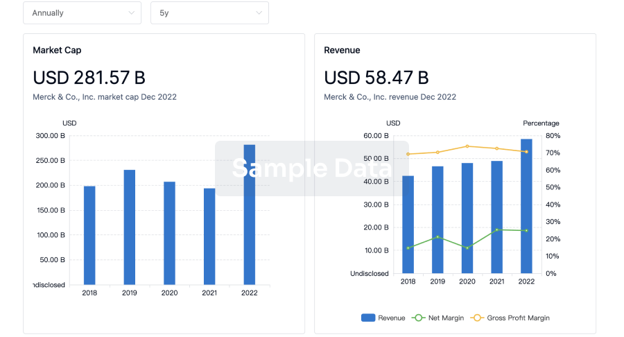
Profit
Explore the financial positions of over 360K organizations with Synapse.
login
or
Grant & Funding(NIH)
Access more than 2 million grant and funding information to elevate your research journey.
login
or
Investment
Gain insights on the latest company investments from start-ups to established corporations.
login
or
Financing
Unearth financing trends to validate and advance investment opportunities.
login
or
AI Agents Built for Biopharma Breakthroughs
Accelerate discovery. Empower decisions. Transform outcomes.
Get started for free today!
Accelerate Strategic R&D decision making with Synapse, PatSnap’s AI-powered Connected Innovation Intelligence Platform Built for Life Sciences Professionals.
Start your data trial now!
Synapse data is also accessible to external entities via APIs or data packages. Empower better decisions with the latest in pharmaceutical intelligence.
Bio
Bio Sequences Search & Analysis
Sign up for free
Chemical
Chemical Structures Search & Analysis
Sign up for free