Request Demo
How to Use Flow Cytometry for T Cell Subset Analysis
28 May 2025
Flow cytometry is a powerful analytical tool widely used in immunology to analyze the phenotypic and functional characteristics of cells, particularly T cell subsets. Its ability to rapidly process thousands of cells per second and provide detailed quantitative and qualitative data makes it indispensable for T cell analysis. This blog will guide you through the process of using flow cytometry for T cell subset analysis, from sample preparation to data interpretation.
Understanding T Cell Subsets
T cells are a vital component of the adaptive immune system, and they can be divided into several subsets, each with distinct functions. The main T cell subsets include CD4+ T helper cells, CD8+ cytotoxic T cells, regulatory T cells (Tregs), and memory T cells. CD4+ T cells help orchestrate the immune response by activating other immune cells, whereas CD8+ T cells directly kill infected or malignant cells. Tregs play a crucial role in maintaining immune tolerance and preventing autoimmune diseases, while memory T cells ensure a rapid and robust response upon re-exposure to antigens.
Preparing Samples for Flow Cytometry
Sample preparation is a critical step in flow cytometry and can significantly affect the accuracy and reliability of your results. Start by obtaining a single-cell suspension from your biological sample, such as peripheral blood, lymph nodes, or spleen. Red blood cell lysis may be necessary, especially when working with blood samples. Once you have a single-cell suspension, count the cells to ensure you have a sufficient number for analysis. Typically, flow cytometry requires a minimum of 100,000 events per sample to obtain reliable data.
Staining Cells with Antibodies
The next step is staining your cells with fluorescently labeled antibodies specific to the surface markers that define your T cell subsets of interest. For example, CD3 is a pan-T cell marker, CD4 is used to identify helper T cells, and CD8 marks cytotoxic T cells. Additional markers such as CD25 and FOXP3 can be used to identify regulatory T cells, while markers like CD45RO and CCR7 can help differentiate memory T cell subsets.
It is crucial to optimize antibody concentrations and staining conditions to minimize non-specific binding and maximize signal specificity. Proper controls are also essential for accurate data interpretation, including unstained cells, single-stained controls for each fluorochrome, and isotype controls.
Running the Flow Cytometer
Once your cells are stained and ready, it's time to run your samples on the flow cytometer. Make sure that your instrument is properly calibrated, and the compensation settings are adjusted to account for spectral overlap between different fluorochromes. Begin by running your experimental controls to establish the correct gating strategy. This process helps to define the positive and negative populations for each marker and set the gates to accurately quantify each T cell subset.
Analyzing Flow Cytometry Data
Data analysis is a key component of flow cytometry, and it involves interpreting the complex datasets generated by the instrument. Use flow cytometry analysis software to create dot plots and histograms, which help visualize the distribution of cell populations based on their fluorescence intensity. Gating strategies are crucial for defining and quantifying the T cell subsets of interest. Start by gating on the lymphocyte population using forward and side scatter, then refine your gating to identify CD3+ T cells, and subsequently, their CD4+ and CD8+ subsets. Further gating can be applied to identify additional subsets like Tregs or memory T cells based on specific marker expression.
Interpreting and Presenting Results
Once your data is analyzed, the final step is interpreting and presenting your results. Consider the biological context and the experimental question you are addressing. Look for changes in the proportion or absolute number of T cell subsets and assess their relevance to the physiological or pathological condition being studied. When presenting your data, clear and concise visualization, such as bar graphs or pie charts, can effectively communicate your findings to your audience.
In conclusion, flow cytometry is an invaluable tool for analyzing T cell subsets, offering deep insights into their roles and functions in the immune system. By following the steps outlined in this blog, you can effectively use flow cytometry to explore the complex dynamics of T cells in health and disease. Whether you are studying immune responses to infections, cancer, or autoimmune conditions, mastering flow cytometry will enhance your research capabilities and contribute to advancing our understanding of immunology.
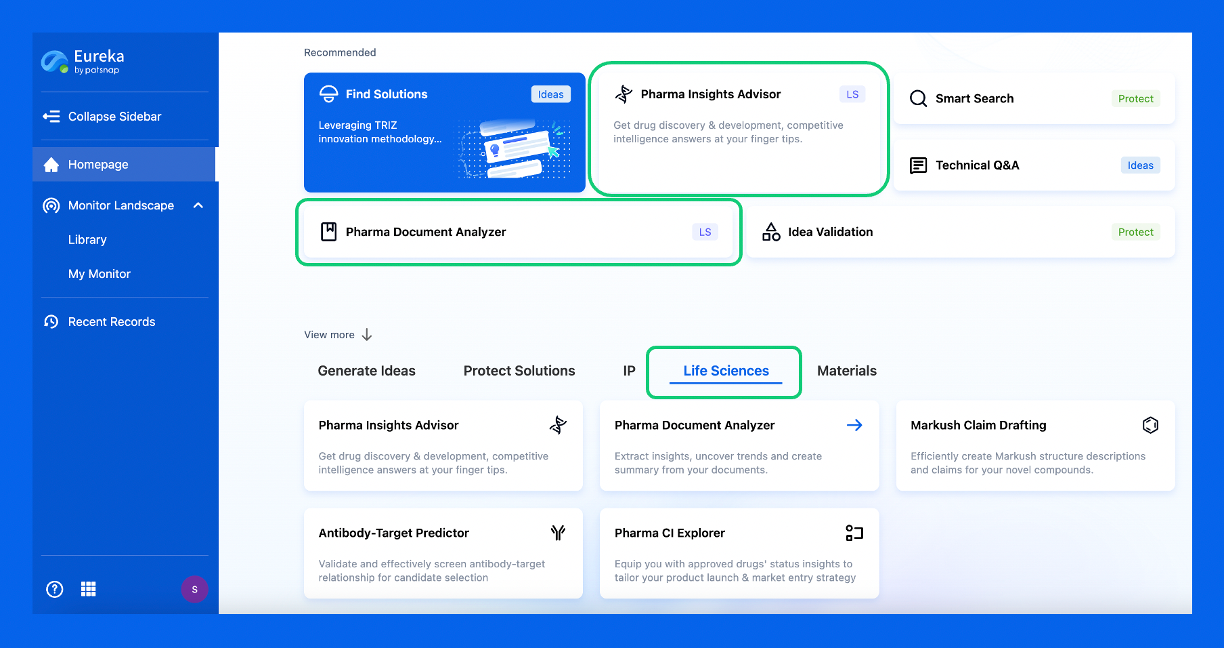
Discover Eureka LS: AI Agents Built for Biopharma Efficiency
Stop wasting time on biopharma busywork. Meet Eureka LS - your AI agent squad for drug discovery.
▶ See how 50+ research teams saved 300+ hours/month
From reducing screening time to simplifying Markush drafting, our AI Agents are ready to deliver immediate value. Explore Eureka LS today and unlock powerful capabilities that help you innovate with confidence.

AI Agents Built for Biopharma Breakthroughs
Accelerate discovery. Empower decisions. Transform outcomes.
Get started for free today!
Accelerate Strategic R&D decision making with Synapse, PatSnap’s AI-powered Connected Innovation Intelligence Platform Built for Life Sciences Professionals.
Start your data trial now!
Synapse data is also accessible to external entities via APIs or data packages. Empower better decisions with the latest in pharmaceutical intelligence.