Request Demo
What is the mechanism of Flortaucipir F-18?
17 July 2024
Flortaucipir F-18 is a radiopharmaceutical agent used in positron emission tomography (PET) imaging to visualize tau protein depositions in the brain, a hallmark feature of Alzheimer’s disease and other tauopathies. The mechanism through which Flortaucipir F-18 operates is both intricate and fascinating, revolving around its ability to bind selectively to tau proteins, allowing for the visualization of pathological changes within the brain.
First and foremost, Flortaucipir F-18 is a fluorine-18 labeled tracer. Fluorine-18 is a radioisotope that emits positrons, making it ideal for PET imaging due to its relatively short half-life of approximately 110 minutes, which minimizes radiation exposure while still providing sufficient time for imaging procedures. The incorporation of fluorine-18 into the molecular structure of Flortaucipir ensures that the tracer can be detected with high sensitivity during PET scans.
The core component of Flortaucipir F-18's mechanism is its specific binding affinity for misfolded tau proteins. Tau proteins are associated with microtubules in neurons, playing a crucial role in maintaining cell structure and function. In Alzheimer's disease and other tauopathies, tau proteins become hyperphosphorylated and misfold, leading to the formation of neurofibrillary tangles. These tangles disrupt normal cellular function and contribute to neuronal death.
Flortaucipir F-18 is designed to cross the blood-brain barrier efficiently, a critical requirement for any neuroimaging agent. Once in the brain, it binds with high affinity to the beta-sheet structures present in the misfolded tau proteins. This high specificity ensures that the tracer provides an accurate representation of tau pathology, distinguishing it from other potential sources of protein aggregation such as amyloid plaques.
Upon administration, Flortaucipir F-18 is injected intravenously and circulates through the bloodstream, eventually crossing into the central nervous system. The tracer's binding to tau deposits allows it to "light up" these areas during a PET scan. The emitted positrons from Fluorine-18 undergo annihilation upon encountering electrons, producing gamma photons that are detected by the PET scanner. This detection process generates detailed images that highlight regions of the brain with high tau burden, providing clinicians with valuable insights into the extent and distribution of tau pathology.
The quantitative nature of PET imaging with Flortaucipir F-18 allows for the assessment of disease progression and the evaluation of therapeutic interventions targeting tau pathology. By providing a non-invasive method to visualize tau depositions, Flortaucipir F-18 facilitates early diagnosis and monitoring of Alzheimer's disease, potentially improving patient outcomes through timely and targeted treatment strategies.
In summary, the mechanism of Flortaucipir F-18 revolves around its ability to specifically bind to misfolded tau proteins in the brain, allowing for the visualization of tau pathology through PET imaging. Its design leverages the radiolabeling of fluorine-18 to produce detectable signals, providing crucial insights into the presence and progression of neurodegenerative diseases characterized by tau protein aggregation.
How to obtain the latest development progress of all drugs?
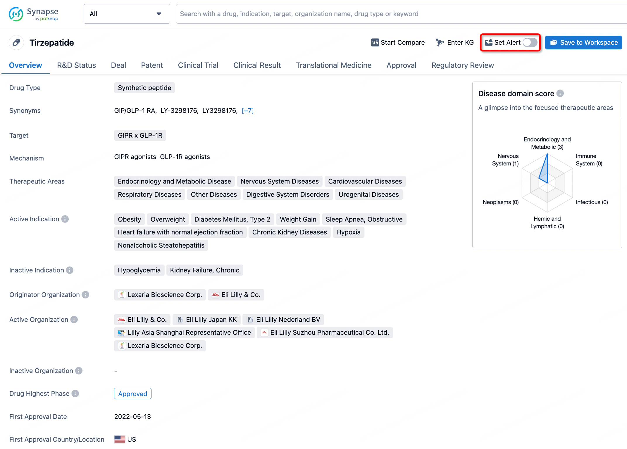
In the Synapse database, you can stay updated on the latest research and development advances of all drugs. This service is accessible anytime and anywhere, with updates available daily or weekly. Use the "Set Alert" function to stay informed. Click on the image below to embark on a brand new journey of drug discovery!


AI Agents Built for Biopharma Breakthroughs
Accelerate discovery. Empower decisions. Transform outcomes.
Get started for free today!
Accelerate Strategic R&D decision making with Synapse, PatSnap’s AI-powered Connected Innovation Intelligence Platform Built for Life Sciences Professionals.
Start your data trial now!
Synapse data is also accessible to external entities via APIs or data packages. Empower better decisions with the latest in pharmaceutical intelligence.