Request Demo
What is the mechanism of Flotufolastat F-18?
17 July 2024
Flotufolastat F-18, also known as ^18F-DCFPyL, is an advanced radiotracer used in positron emission tomography (PET) imaging, primarily to identify and evaluate prostate cancer. Understanding the mechanism of Flotufolastat F-18 involves delving into its biochemical interactions, its role in diagnostic imaging, and its impact on patient management in oncology.
Flotufolastat F-18 is a radiopharmaceutical compound, where the F-18 refers to the radioactive isotope fluorine-18. This isotope is pivotal due to its favorable half-life and positron emission properties, making it ideal for PET imaging. The mechanism of Flotufolastat F-18 centers around its ability to target the prostate-specific membrane antigen (PSMA), a protein highly expressed on the surface of prostate cancer cells.
Upon administration, Flotufolastat F-18 binds specifically to PSMA. This antigen is a glycoprotein found in large quantities on prostate cancer cells, as well as on the neovasculature of other solid tumors, but not significantly present in most normal tissues. The selective binding of Flotufolastat F-18 to PSMA enables precise targeting of cancer cells, which is crucial for both the detection and staging of prostate cancer.
The compound itself consists of two main components: the targeting moiety and the radiolabel. The targeting moiety is responsible for the high affinity binding to the PSMA, and the radiolabel, which is the fluorine-18 isotope, emits positrons detectable by the PET scanner. When Flotufolastat F-18 binds to PSMA-expressing cells, it creates a distinct signal that can be captured via PET imaging.
The biochemical interaction begins at the molecular level where the targeting moiety of Flotufolastat F-18 binds to the extracellular domain of PSMA. Once bound, the complex is internalized into the cancer cell. The fluorine-18 isotope, due to its radioactive decay, emits positrons. When these positrons encounter electrons within the body's tissues, they annihilate each other, producing gamma photons. These gamma photons are detected by the PET scanner, creating detailed images of areas with high PSMA expression.
The images produced by Flotufolastat F-18 PET scans are highly sensitive and specific. This allows for accurate localization of primary and metastatic prostate cancer lesions. The precise imaging aids clinicians in determining the extent of the disease, evaluating the effectiveness of ongoing treatments, and planning appropriate therapeutic interventions.
Moreover, the high affinity and specificity of Flotufolastat F-18 for PSMA provide a significant advantage over other imaging modalities. Its ability to highlight even small and otherwise undetectable lesions means that it can identify cancer spread at an earlier stage, potentially leading to better patient outcomes through early intervention.
In summary, the mechanism of Flotufolastat F-18 involves its targeted binding to PSMA, internalization into cancer cells, and the subsequent emission of detectable positrons. This enables highly accurate PET imaging for the detection and management of prostate cancer. The selective nature of this radiotracer ensures that clinicians can make more informed decisions, ultimately enhancing patient care in oncology.
How to obtain the latest development progress of all drugs?
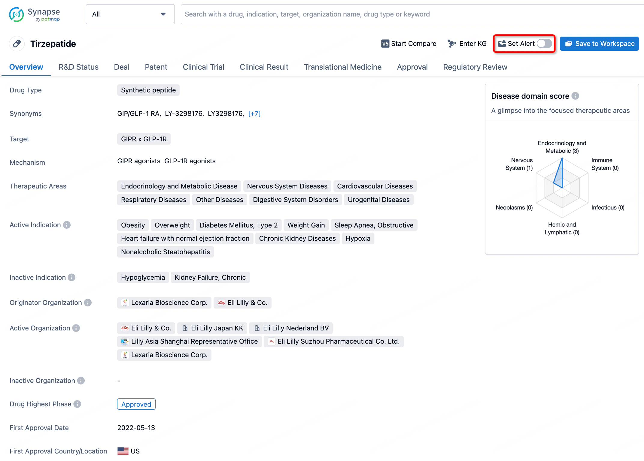
In the Synapse database, you can stay updated on the latest research and development advances of all drugs. This service is accessible anytime and anywhere, with updates available daily or weekly. Use the "Set Alert" function to stay informed. Click on the image below to embark on a brand new journey of drug discovery!


AI Agents Built for Biopharma Breakthroughs
Accelerate discovery. Empower decisions. Transform outcomes.
Get started for free today!
Accelerate Strategic R&D decision making with Synapse, PatSnap’s AI-powered Connected Innovation Intelligence Platform Built for Life Sciences Professionals.
Start your data trial now!
Synapse data is also accessible to external entities via APIs or data packages. Empower better decisions with the latest in pharmaceutical intelligence.