Request Demo
What is the mechanism of Flutemetamol F-18?
17 July 2024
Flutemetamol F-18 is a radiopharmaceutical used primarily for positron emission tomography (PET) imaging of the brain to detect beta-amyloid plaques, which are associated with Alzheimer's disease. Understanding the mechanism of Flutemetamol F-18 involves delving into its biochemical properties, its interaction with biological systems, and its role in medical diagnostics.
At its core, Flutemetamol F-18 contains a fluorine-18 isotope, a positron-emitting radionuclide. Fluorine-18 has a relatively short half-life of about 110 minutes, which makes it suitable for PET imaging due to its ability to provide high-resolution images while limiting the radiation exposure to the patient.
The mechanism begins with the preparation of the compound. Flutemetamol is a synthetic analog of thioflavin T, a dye that binds specifically to amyloid fibrils. The fluorine-18 isotope is incorporated into the molecular structure of flutemetamol through a nucleophilic substitution reaction, resulting in Flutemetamol F-18. This radiolabeled compound retains the amyloid-binding properties of thioflavin T, but with the added benefit of being detectable via PET imaging.
Once administered intravenously, Flutemetamol F-18 circulates through the bloodstream and crosses the blood-brain barrier. It has a high affinity for beta-amyloid plaques, which are insoluble protein aggregates that form in the brains of individuals with Alzheimer's disease. The compound selectively binds to these plaques due to its chemical structure, which interacts with the beta-sheet conformation of amyloid fibrils.
After binding to the beta-amyloid plaques, the fluorine-18 isotope emits positrons as it undergoes radioactive decay. These positrons collide with electrons in the brain tissue, resulting in the emission of gamma photons. The PET scanner detects these gamma photons, and a computer reconstructs the data into a detailed image showing the distribution of Flutemetamol F-18 in the brain.
The intensity of the signal in the PET images correlates with the density of beta-amyloid plaques. Higher uptake of Flutemetamol F-18 indicates a greater presence of amyloid plaques, which is a hallmark of Alzheimer's disease. Clinicians can use this information to support a diagnosis of Alzheimer's disease or to assess the extent of amyloid pathology in the brain.
Flutemetamol F-18 is particularly valuable because it enables early detection of beta-amyloid plaques, even before the onset of clinical symptoms. This early detection can be crucial for initiating timely interventions and for enrolling patients in clinical trials aimed at slowing the progression of Alzheimer's disease.
Moreover, Flutemetamol F-18 PET imaging can be used to monitor the effectiveness of therapeutic interventions targeting amyloid plaques. By comparing images taken before and after treatment, clinicians can assess whether a given therapy is reducing the amyloid burden in the brain.
In summary, the mechanism of Flutemetamol F-18 involves its synthesis as a fluorine-18-labeled analog of thioflavin T, its selective binding to beta-amyloid plaques in the brain, and the subsequent detection of emitted gamma photons through PET imaging. This process allows for the non-invasive visualization of amyloid pathology, providing valuable insights into the diagnosis and management of Alzheimer's disease.
How to obtain the latest development progress of all drugs?
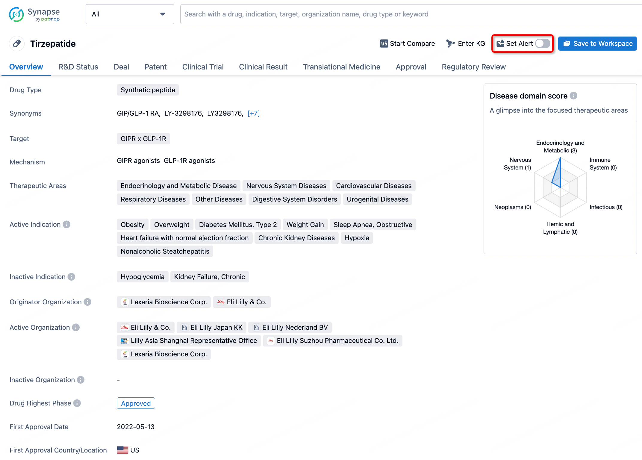
In the Synapse database, you can stay updated on the latest research and development advances of all drugs. This service is accessible anytime and anywhere, with updates available daily or weekly. Use the "Set Alert" function to stay informed. Click on the image below to embark on a brand new journey of drug discovery!


AI Agents Built for Biopharma Breakthroughs
Accelerate discovery. Empower decisions. Transform outcomes.
Get started for free today!
Accelerate Strategic R&D decision making with Synapse, PatSnap’s AI-powered Connected Innovation Intelligence Platform Built for Life Sciences Professionals.
Start your data trial now!
Synapse data is also accessible to external entities via APIs or data packages. Empower better decisions with the latest in pharmaceutical intelligence.