Request Demo
What is the mechanism of Gadoteridol?
17 July 2024
Gadoteridol is a gadolinium-based contrast agent (GBCA) widely used in magnetic resonance imaging (MRI) to enhance the quality of the images. Understanding the mechanism of action of Gadoteridol is crucial for appreciating its efficacy and safety profile.
Gadoteridol is a non-ionic, macrocyclic contrast agent. Its primary component is gadolinium (Gd), a rare earth metal that has paramagnetic properties. These properties make gadolinium particularly useful in MRI, as it can enhance the relaxation rates of water protons in the body, leading to an improvement in image contrast.
When Gadoteridol is administered intravenously, it circulates through the bloodstream and distributes throughout the extracellular fluid space. The mechanism by which it enhances MRI images can be broken down into several key phases:
1. **Distribution in the Body**:
Upon entering the bloodstream, Gadoteridol diffuses through the extracellular space. Because it is a low-molecular-weight agent, it can easily permeate tissues and organs, except for those protected by the blood-brain barrier, unless the barrier is compromised.
2. **Relaxation Enhancement**:
The primary purpose of Gadoteridol is to enhance the contrast of MRI images by altering the magnetic properties of nearby water molecules. The gadolinium ions in Gadoteridol have seven unpaired electrons, which create a significant magnetic moment. When placed in an external magnetic field, such as that of an MRI scanner, these gadolinium ions interact with the hydrogen nuclei (protons) in water molecules.
This interaction shortens the T1 and T2 relaxation times of the protons. T1 relaxation time is the time it takes for spinning protons in tissue to realign with the magnetic field after being knocked out of alignment. T2 relaxation time is the time it takes for the protons to lose phase coherence among the spinning protons. By shortening these relaxation times, Gadoteridol makes the affected tissues appear brighter on T1-weighted MRI images and can also affect T2-weighted images, though to a lesser extent.
3. **Imaging Phase**:
Once Gadoteridol has been distributed and has altered the relaxation times in the target tissues, MRI sequences can be optimized to take advantage of these changes. T1-weighted sequences are typically used to best visualize the areas where Gadoteridol has accumulated, thereby providing enhanced contrast between different tissues, making abnormalities like tumors, inflammation, or vascular lesions more conspicuous.
4. **Excretion**:
Gadoteridol is mainly excreted through the kidneys via glomerular filtration. It has a relatively rapid elimination half-life, typically a few hours in patients with normal renal function. This rapid clearance helps minimize the potential for toxicity and reduces the duration of any possible side effects.
5. **Safety Profile**:
The macrocyclic structure of Gadoteridol is designed to tightly bind the gadolinium ion, reducing the risk of free gadolinium release, which can be toxic. This structural stability is advantageous over some linear GBCAs, which are more prone to releasing gadolinium. As a result, Gadoteridol has a favorable safety profile, with a low incidence of adverse effects.
In summary, the mechanism of Gadoteridol involves its distribution in the extracellular space, enhancement of proton relaxation times, optimized MRI imaging sequences, and rapid renal excretion. These combined actions make Gadoteridol an effective and safe contrast agent for enhancing the quality and diagnostic utility of MRI scans. Understanding these mechanisms helps clinicians optimally use Gadoteridol to achieve the best possible imaging outcomes.
How to obtain the latest development progress of all drugs?
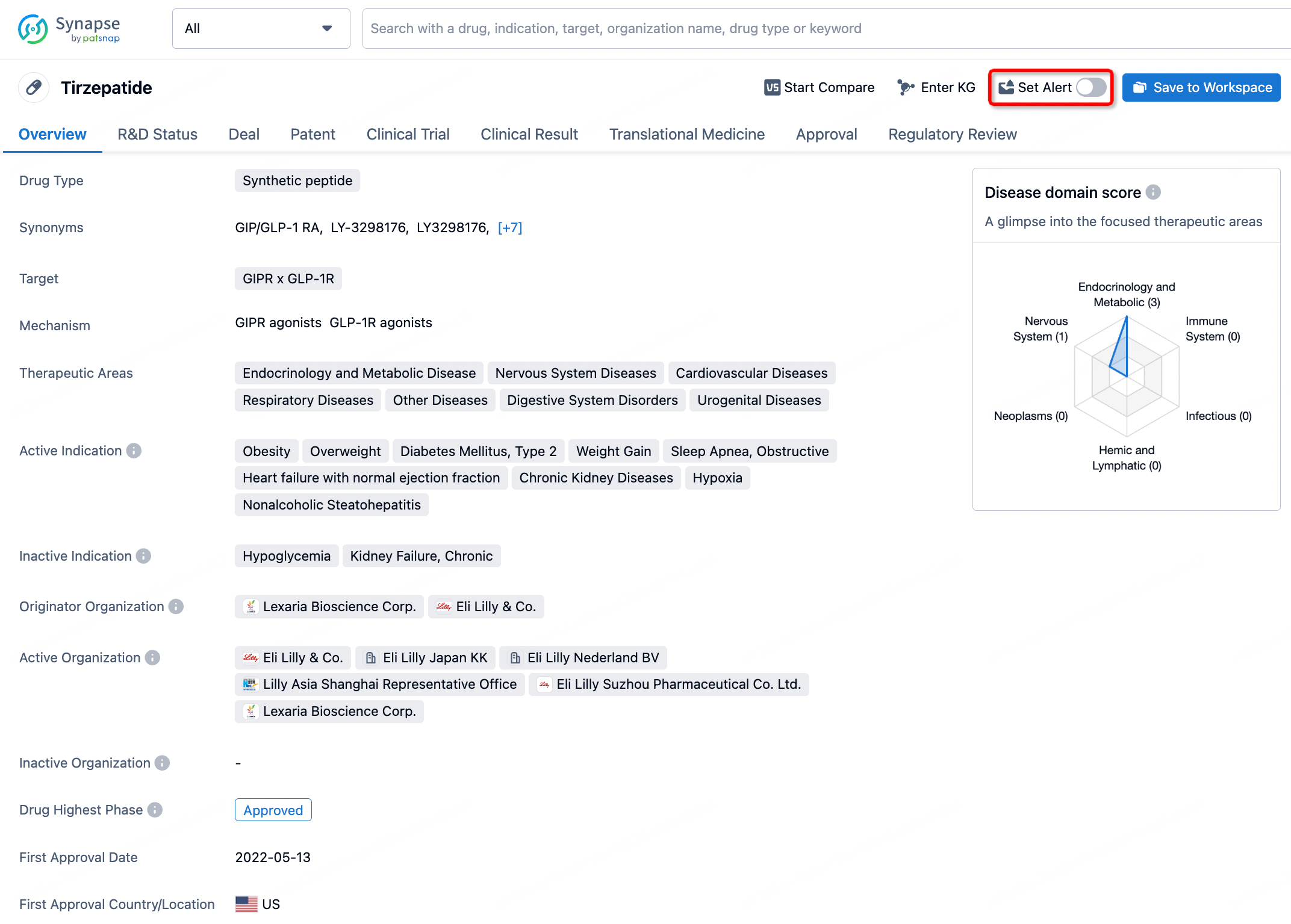
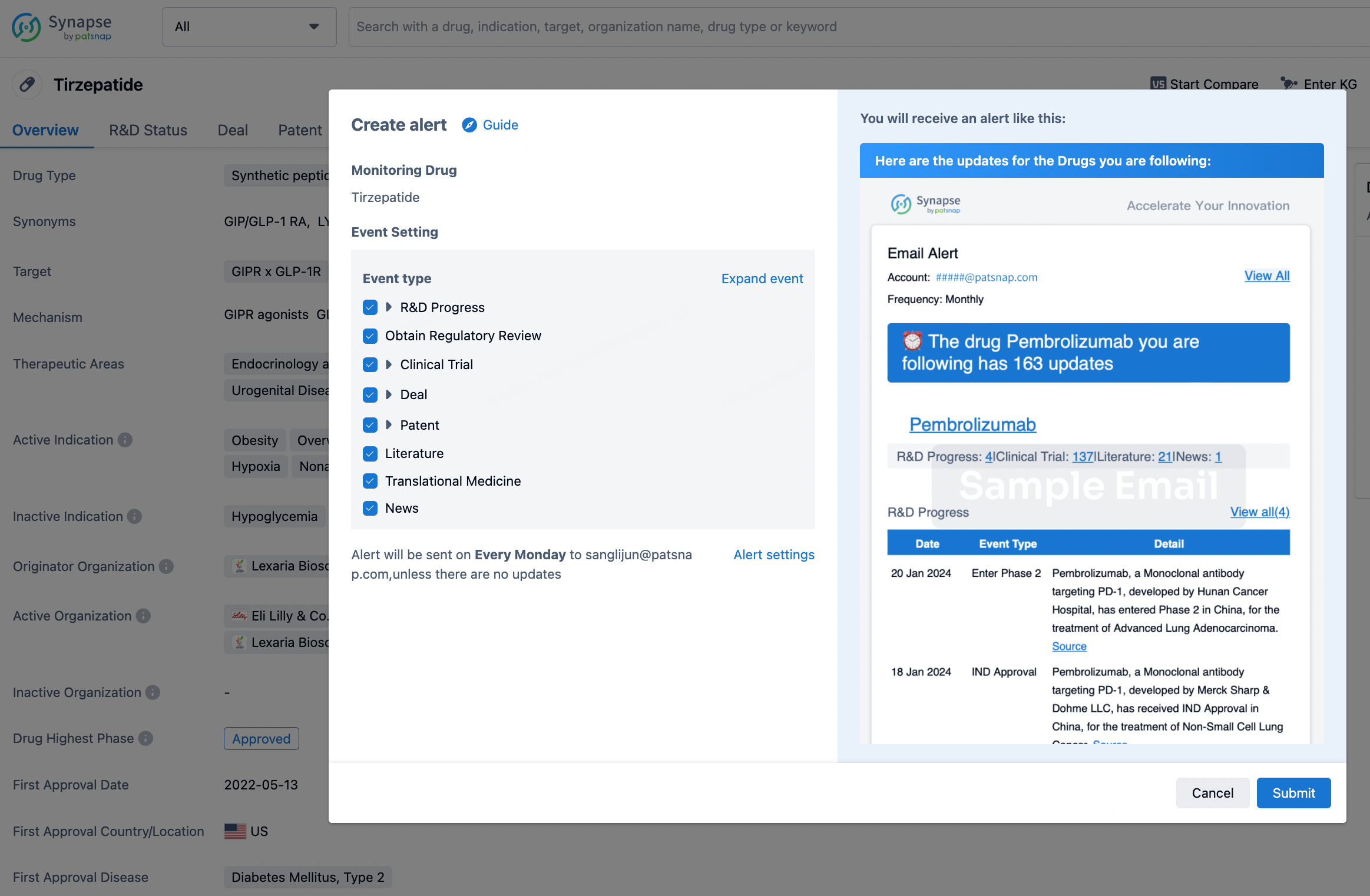
In the Synapse database, you can stay updated on the latest research and development advances of all drugs. This service is accessible anytime and anywhere, with updates available daily or weekly. Use the "Set Alert" function to stay informed. Click on the image below to embark on a brand new journey of drug discovery!
AI Agents Built for Biopharma Breakthroughs
Accelerate discovery. Empower decisions. Transform outcomes.
Get started for free today!
Accelerate Strategic R&D decision making with Synapse, PatSnap’s AI-powered Connected Innovation Intelligence Platform Built for Life Sciences Professionals.
Start your data trial now!
Synapse data is also accessible to external entities via APIs or data packages. Empower better decisions with the latest in pharmaceutical intelligence.


