Request Demo
What is the mechanism of Iopamidol?
17 July 2024
Iopamidol is a non-ionic, water-soluble radiographic contrast agent widely used in medical imaging to enhance the visibility of internal structures during radiographic examinations. Understanding the mechanism of iopamidol involves exploring its chemical composition, pharmacokinetics, and the way it interacts with biological tissues during imaging procedures.
Iopamidol is a triiodinated benzamide derivative, which contributes significantly to its radiopacity. The iodine atoms present in the molecule are pivotal as they absorb X-rays more effectively than the surrounding tissues, thereby providing a contrast that delineates structures more clearly on radiographic films or digital images. This improved contrast is essential for accurately diagnosing and evaluating various medical conditions.
The pharmacokinetics of iopamidol reveal that, upon intravenous administration, the agent rapidly disperses into the bloodstream and is distributed throughout the body's extracellular fluid. It does not bind significantly to plasma proteins, allowing for a relatively quick circulation and subsequent excretion. The renal system plays a crucial role in the elimination process, with the majority of iopamidol being excreted unchanged via the kidneys within 24 hours of administration. This rapid clearance minimizes the risk of prolonged exposure and potential toxicity.
During imaging procedures, iopamidol's mechanism of action is relatively straightforward yet highly effective. When administered, it traverses the vascular system and accumulates in areas with high extracellular fluid content, such as blood vessels, the urinary tract, and soft tissues. The presence of the iodine atoms within the iopamidol molecules creates a differential absorption of X-rays, resulting in enhanced contrast of the targeted areas. This contrast enhances the visibility of anatomical structures and pathological changes, aiding radiologists and medical professionals in making accurate diagnoses.
Iopamidol’s efficacy is also influenced by its osmolality and viscosity. Non-ionic contrast agents like iopamidol are designed to have lower osmolality compared to older ionic agents, reducing the incidence of adverse reactions such as pain at the injection site, nausea, and cardiovascular disturbances. Its lower osmolality ensures better patient tolerance and reduces the risk of complications during and after the imaging procedure.
Safety profiles of iopamidol are well-documented, with a majority of patients tolerating the agent well. However, like all contrast media, it is not devoid of risks. Allergic reactions, although rare, can occur, and pre-existing conditions such as renal impairment may necessitate cautious use or alternative imaging strategies. Pre-screening patients for risk factors and ensuring adequate hydration are essential steps in minimizing potential adverse effects.
In summary, iopamidol functions as an effective radiographic contrast agent through its iodine-based molecular structure, rapid pharmacokinetics, and favorable safety profile. Its ability to enhance the visibility of internal structures during imaging procedures plays a crucial role in the accurate diagnosis and management of various medical conditions, underscoring its importance in modern medical imaging practices.
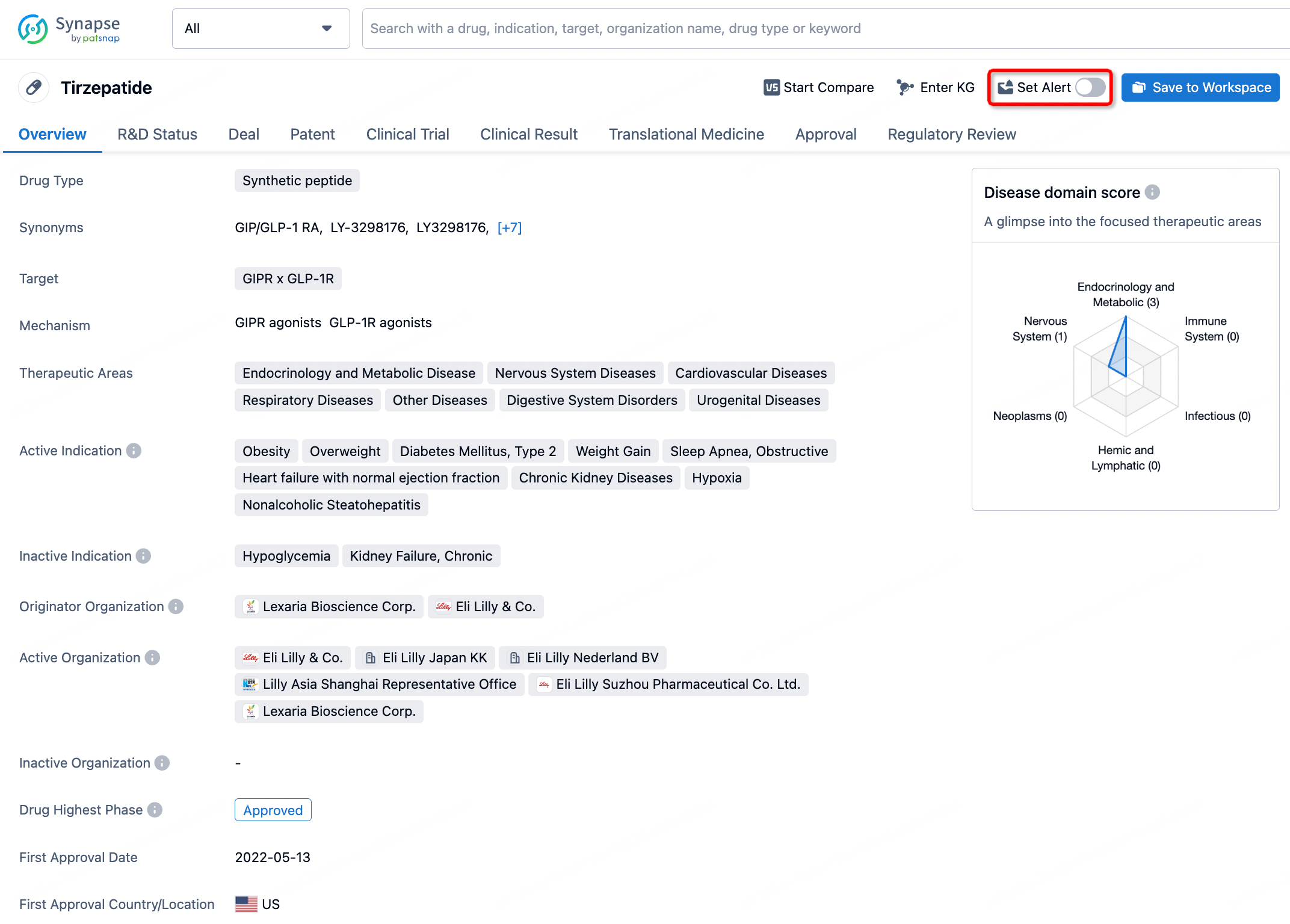
How to obtain the latest development progress of all drugs?
In the Synapse database, you can stay updated on the latest research and development advances of all drugs. This service is accessible anytime and anywhere, with updates available daily or weekly. Use the "Set Alert" function to stay informed. Click on the image below to embark on a brand new journey of drug discovery!


AI Agents Built for Biopharma Breakthroughs
Accelerate discovery. Empower decisions. Transform outcomes.
Get started for free today!
Accelerate Strategic R&D decision making with Synapse, PatSnap’s AI-powered Connected Innovation Intelligence Platform Built for Life Sciences Professionals.
Start your data trial now!
Synapse data is also accessible to external entities via APIs or data packages. Empower better decisions with the latest in pharmaceutical intelligence.