Request Demo
What is the mechanism of Ioxilan?
18 July 2024
Ioxilan is a nonionic, low-osmolar contrast agent used extensively in radiographic imaging procedures such as computed tomography (CT) scans, angiography, and urography. Its primary function is to enhance the contrast of images obtained during these procedures, allowing for a more detailed and accurate visualization of bodily structures and abnormalities. Understanding the mechanism of ioxilan involves delving into its chemical properties, pharmacokinetics, and the physiological interactions that make it effective as a contrast medium.
Chemically, ioxilan belongs to the class of iodinated contrast agents. It is a triiodinated, benzene derivative with the chemical formula C18H24I3N3O8. The iodine atoms are critical to its function, as their high atomic number and electron density significantly increase the X-ray absorption of the areas where ioxilan is present. This results in the enhanced contrast required for detailed imaging.
Upon administration, ioxilan is typically injected intravenously or intra-arterially, depending on the type of imaging procedure. Its low osmolarity compared to older, high-osmolar contrast agents means it has a lower risk of causing adverse reactions such as nephrotoxicity or osmotically induced vascular and extravascular fluid shifts. This makes it a safer option for patients, particularly those with pre-existing renal conditions or other comorbidities.
Pharmacokinetically, ioxilan is rapidly distributed throughout the extracellular fluid space after administration. Its distribution is predominantly influenced by blood flow to various tissues, with high perfusion organs like the kidneys, liver, and brain receiving the contrast agent more quickly. The contrast agent does not undergo significant metabolism and is primarily excreted unchanged by the kidneys through glomerular filtration. This efficient renal clearance ensures that ioxilan does not remain in the body for extended periods, reducing the likelihood of prolonged exposure and potential toxicity.
During imaging procedures, ioxilan's high iodine content absorbs the X-rays more effectively than the surrounding tissues. This differential absorption creates a distinct contrast on the radiographic film or digital detector, highlighting structures such as blood vessels, organs, and any pathological lesions. In vascular imaging, for instance, ioxilan can clearly delineate arterial and venous anatomy, aiding in the diagnosis of conditions like aneurysms, stenoses, and occlusions.
The safety profile of ioxilan is another crucial aspect of its mechanism. Its low osmolarity translates to fewer adverse effects such as pain, heat sensation, or local tissue irritation at the injection site. Additionally, nonionic contrast agents like ioxilan are less likely to induce allergic reactions compared to their ionic counterparts. However, it is still essential to conduct a thorough patient history and assessment, as some individuals may still experience hypersensitivity reactions or other side effects.
In summary, the mechanism of ioxilan as a contrast agent in radiographic imaging is anchored in its chemical structure, pharmacokinetics, and interaction with X-rays. Its iodine atoms enhance X-ray absorption, while its low osmolarity and rapid renal clearance contribute to its safety and efficacy. Understanding these mechanisms allows healthcare professionals to utilize ioxilan effectively, ensuring high-quality diagnostic imaging with minimal risk to patients.
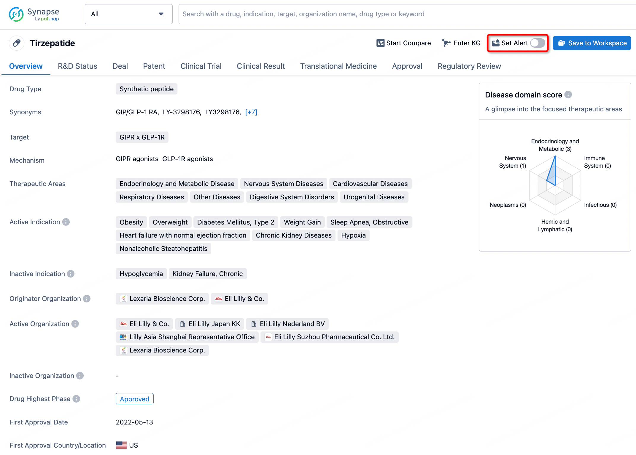
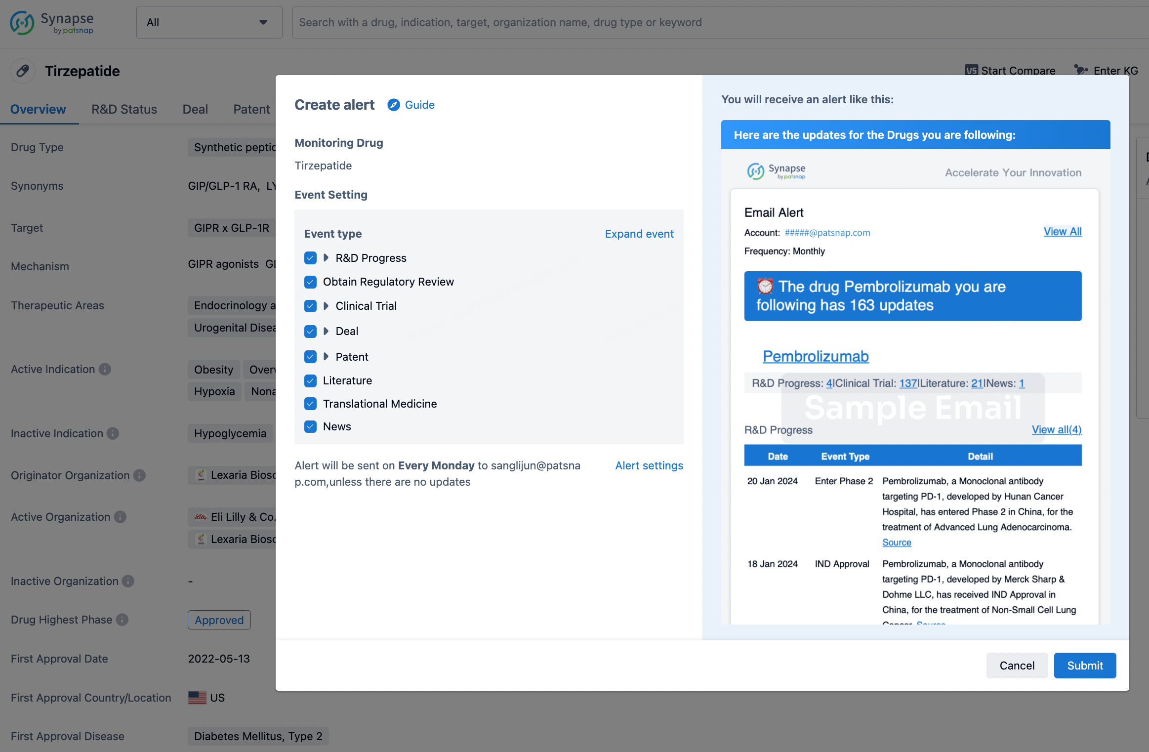
How to obtain the latest development progress of all drugs?
In the Synapse database, you can stay updated on the latest research and development advances of all drugs. This service is accessible anytime and anywhere, with updates available daily or weekly. Use the "Set Alert" function to stay informed. Click on the image below to embark on a brand new journey of drug discovery!
AI Agents Built for Biopharma Breakthroughs
Accelerate discovery. Empower decisions. Transform outcomes.
Get started for free today!
Accelerate Strategic R&D decision making with Synapse, PatSnap’s AI-powered Connected Innovation Intelligence Platform Built for Life Sciences Professionals.
Start your data trial now!
Synapse data is also accessible to external entities via APIs or data packages. Empower better decisions with the latest in pharmaceutical intelligence.