Request Demo
What is the mechanism of Trypsin?
18 July 2024
Trypsin is a serine protease enzyme that plays a crucial role in the digestive system. It is produced in the pancreas as an inactive precursor, trypsinogen, and is then secreted into the small intestine. Here, it is activated to its functional form, trypsin, by the enzyme enteropeptidase. Trypsin is essential for the breakdown of proteins into smaller peptides, which can then be further digested by other proteases into amino acids, the building blocks of life. Understanding the mechanism of trypsin involves delving into its structure, activation process, substrate specificity, and catalytic action.
Structurally, trypsin is a globular protein with a well-defined active site. The active site contains a catalytic triad formed by three amino acids: serine, histidine, and aspartate. These three residues work together to cleave peptide bonds in proteins. The serine residue acts as a nucleophile, the histidine residue functions as a base, and the aspartate stabilizes the histidine. This triad is a hallmark of serine proteases and is crucial for their catalytic activity.
The activation of trypsinogen to trypsin is a finely tuned process that ensures the enzyme is only activated when it reaches the small intestine. Enteropeptidase, an enzyme located on the brush border of the duodenum, recognizes and cleaves a specific peptide bond in trypsinogen, converting it into active trypsin. Once activated, trypsin can also catalyze the conversion of more trypsinogen to trypsin, a process known as autocatalysis. This rapid activation ensures that a sufficient amount of active enzyme is available for protein digestion.
Trypsin exhibits substrate specificity, meaning it selectively cleaves peptide bonds at the carboxyl side of lysine and arginine residues. This specificity is dictated by the structure of the enzyme's active site, which includes a deep pocket that accommodates the side chains of lysine and arginine. When a protein substrate enters the active site, the catalytic triad works in concert to cleave the peptide bond. The serine residue attacks the carbonyl carbon of the peptide bond, forming a tetrahedral intermediate. This intermediate is stabilized by the enzyme's oxyanion hole, a structural feature that helps to lower the activation energy of the reaction.
The histidine residue, acting as a base, accepts a proton from the serine residue, facilitating the nucleophilic attack. Concurrently, the aspartate residue stabilizes the positively charged histidine, ensuring the proper orientation of the catalytic triad. The tetrahedral intermediate then collapses, releasing the C-terminal fragment of the substrate and forming an acyl-enzyme intermediate. This intermediate is subsequently hydrolyzed by a water molecule, which is activated by the histidine residue. The result is the release of the N-terminal fragment of the substrate and the regeneration of the active site, ready for another catalytic cycle.
In summary, the mechanism of trypsin involves a series of well-coordinated steps, starting with its activation from trypsinogen by enteropeptidase. The enzyme's catalytic triad plays a central role in cleaving peptide bonds at specific sites within protein substrates. The structural features of trypsin, including its active site and oxyanion hole, are crucial for its function. Through these intricate processes, trypsin efficiently converts dietary proteins into smaller peptides, facilitating their further digestion and absorption in the small intestine. Understanding the mechanism of trypsin not only provides insights into its biological function but also underscores the elegance of enzymatic catalysis in general.
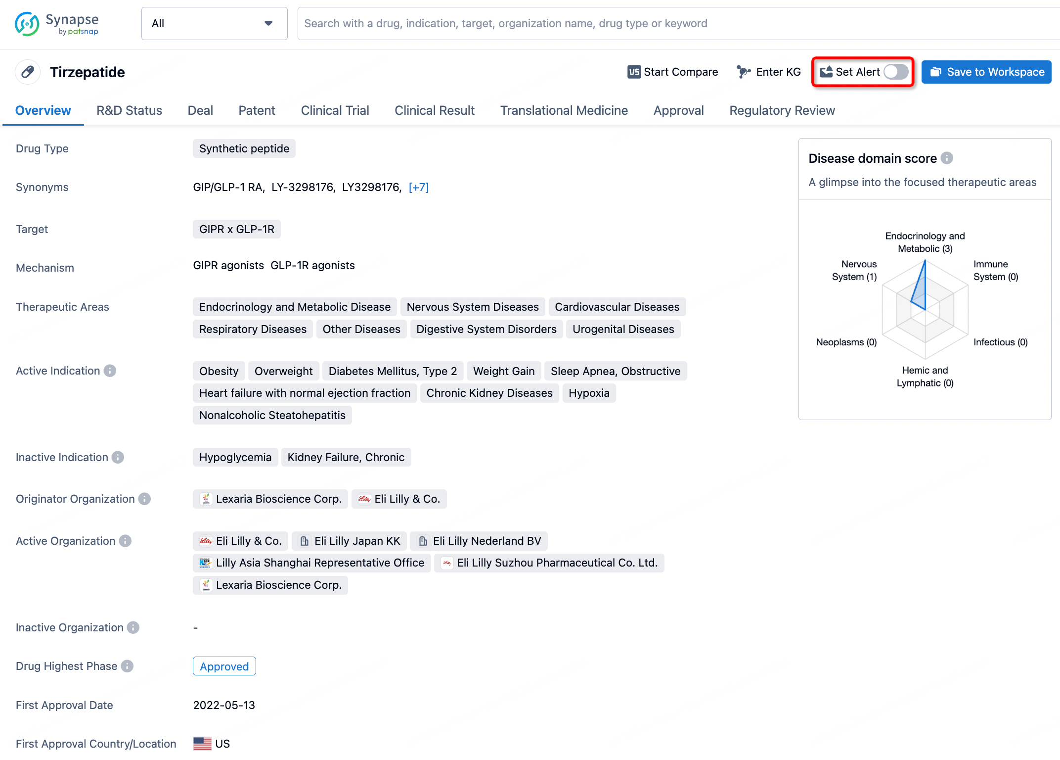
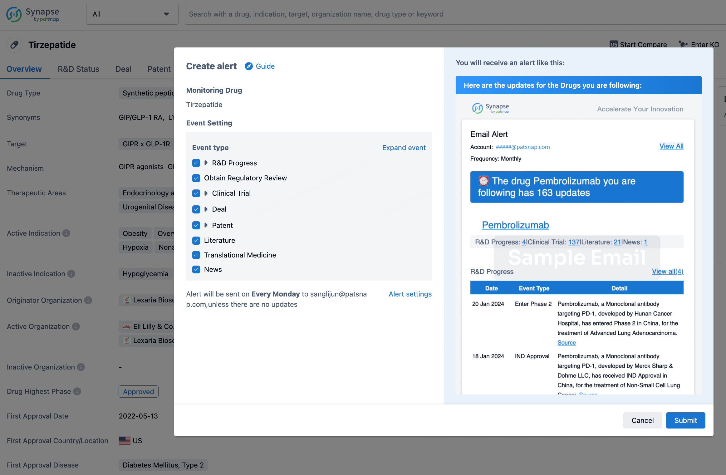
How to obtain the latest development progress of all drugs?
In the Synapse database, you can stay updated on the latest research and development advances of all drugs. This service is accessible anytime and anywhere, with updates available daily or weekly. Use the "Set Alert" function to stay informed. Click on the image below to embark on a brand new journey of drug discovery!

AI Agents Built for Biopharma Breakthroughs
Accelerate discovery. Empower decisions. Transform outcomes.
Get started for free today!
Accelerate Strategic R&D decision making with Synapse, PatSnap’s AI-powered Connected Innovation Intelligence Platform Built for Life Sciences Professionals.
Start your data trial now!
Synapse data is also accessible to external entities via APIs or data packages. Empower better decisions with the latest in pharmaceutical intelligence.