Request Demo
Last update 09 Aug 2025
Caspase-3 inhibitor(Chembridge Research Laboratories)
Last update 09 Aug 2025
Overview
Basic Info
Drug Type Synthetic peptide |
Synonyms- |
Target |
Action inhibitors |
Mechanism caspase 3 inhibitors(Caspase-3 inhibitors) |
Therapeutic Areas- |
Active Indication- |
Inactive Indication- |
Originator Organization |
Active Organization |
Inactive Organization- |
License Organization- |
Drug Highest PhaseDiscovery |
First Approval Date- |
Regulation- |
Structure/Sequence
Molecular FormulaC27H27N5O9 |
InChIKeyHWYYSCJNSUTDTD-ZRELSDDQSA-N |
CAS Registry821800-07-1 |
R&D Status
Login to view more data
Clinical Result
Clinical Result
Indication
Phase
Evaluation
View All Results
Login to view more data
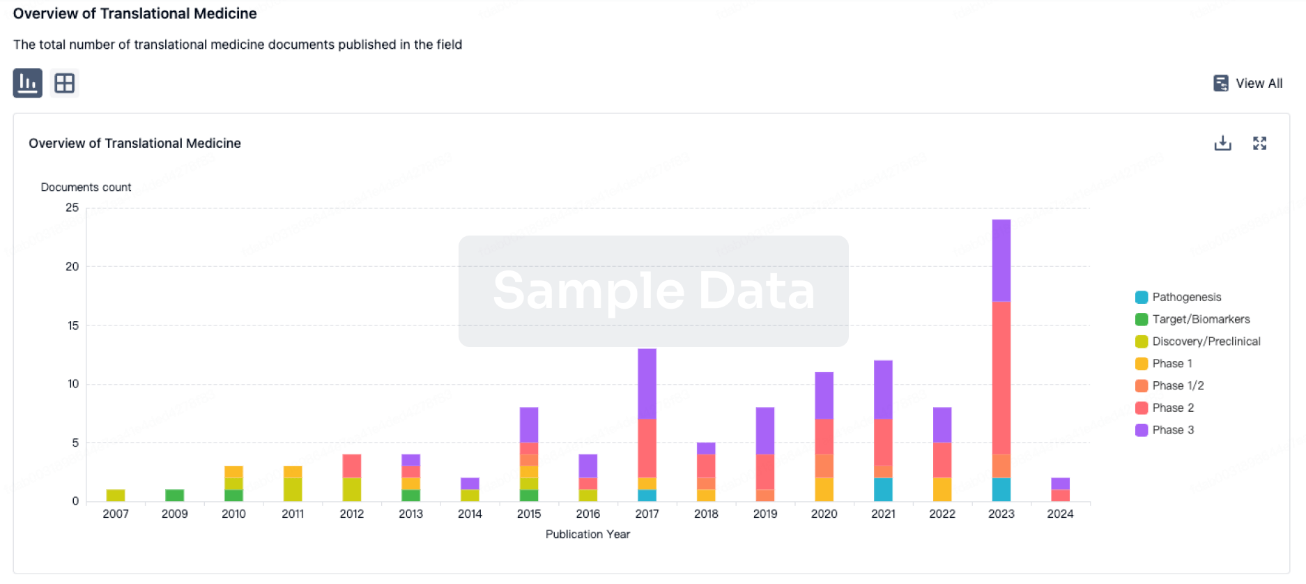
Translational Medicine
Boost your research with our translational medicine data.
login
or
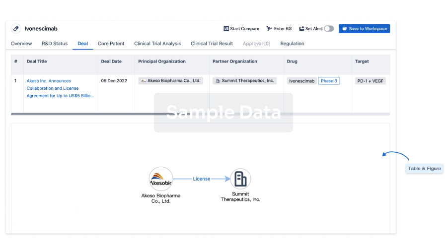
Deal
Boost your decision using our deal data.
login
or
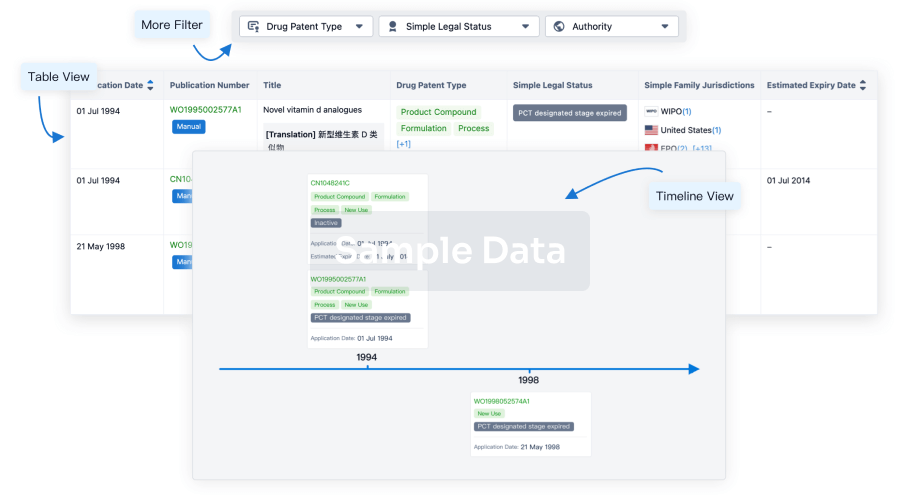
Core Patent
Boost your research with our Core Patent data.
login
or
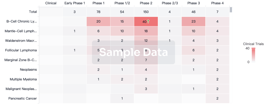
Clinical Trial
Identify the latest clinical trials across global registries.
login
or
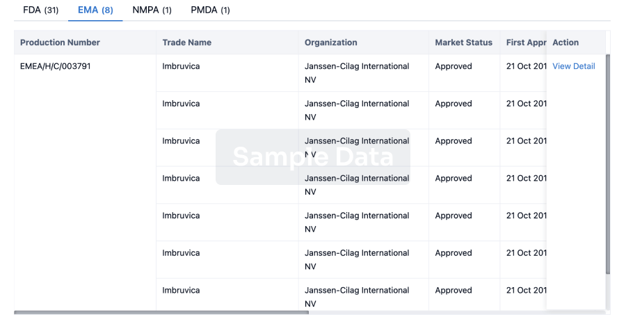
Approval
Accelerate your research with the latest regulatory approval information.
login
or
Biosimilar
Competitive landscape of biosimilars in different countries/locations. Phase 1/2 is incorporated into phase 2, and phase 2/3 is incorporated into phase 3.
login
or

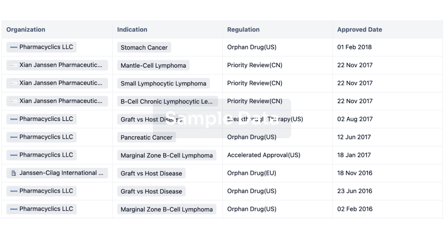
Regulation
Understand key drug designations in just a few clicks with Synapse.
login
or
AI Agents Built for Biopharma Breakthroughs
Accelerate discovery. Empower decisions. Transform outcomes.
Get started for free today!
Accelerate Strategic R&D decision making with Synapse, PatSnap’s AI-powered Connected Innovation Intelligence Platform Built for Life Sciences Professionals.
Start your data trial now!
Synapse data is also accessible to external entities via APIs or data packages. Empower better decisions with the latest in pharmaceutical intelligence.
Bio
Bio Sequences Search & Analysis
Sign up for free
Chemical
Chemical Structures Search & Analysis
Sign up for free