Request Demo
Last update 06 Jul 2025

Antaros Medical AB
Last update 06 Jul 2025
Overview
Tags
Digestive System Disorders
Other Diseases
Immune System Diseases
Diagnostic radiopharmaceuticals
Affibody
Disease domain score
A glimpse into the focused therapeutic areas

Technology Platform
Most used technologies in drug development
Targets
Most frequently developed targets
Related
Target |
Mechanism |
Active Org. |
Originator Org. |
Active Indication |
Inactive Indication |
Drug Highest Phase |
First Approval Ctry. / Loc. |
First Approval Date |
Target |
Mechanism |
Active Org. |
Originator Org. |
Active Indication |
Inactive Indication |
Drug Highest Phase |
First Approval Ctry. / Loc. |
First Approval Date |
NCT06562361
A First-In-Human, Microdosing, Clinical Trial to Investigate Binding of the PET Tracer [68Ga]Ga-DOTA-CYS-ATH001 Targeting PDGFRβ in Healthy Subjects as Compared to Patients With MASH, PSC and CD
NCT06022263
A Prospective, Randomised, Double-blinded, Placebo-controlled Study Investigating the Safety and Tolerability of STA363 in Patients With Radiculopathy Caused by Lumbar Disc Herniation

NCT04307797
A Pilot Study on the Effect of Glucagon and Glucagon-like Peptide-1 Co-agonism on Cardiac Function and Metabolism in Overweight Participants with Type 2 Diabetes (COCONUT)

100 Clinical Results associated with Antaros Medical AB
Login to view more data
Login to view more data
03 Jun 2025SCANDINAVIAN JOURNAL OF GASTROENTEROLOGY
Proteomic signatures for fibrosis in MASLD: a biopsy-proven dual-cohort study
Article
Author: Rorsman, Fredrik ; Gabrysch, Katja ; Hockings, Paul ; Nasr, Patrik ; Eriksson, Niclas ; Hulthe, Johannes ; Vessby, Johan ; Kechagias, Stergios ; Fridén, Michael ; Blomdahl, Julia ; Ekstedt, Mattias ; Ahlström, Håkan ; Risérus, Ulf ; Åberg, Mikael
OBJECTIVES:
Predicting disease progression in metabolic dysfunction-associated steatotic liver disease (MASLD) is challenging, and current non-invasive tests (NITs) lack the precision to replace liver biopsy. This study aimed to identify plasma biomarkers for different stages of fibrosis using affinity-based proteomics in two biopsy-proven cohorts. The primary objective was to identify biomarkers capable of distinguishing between low-to-no fibrosis (F0-1) and significant fibrosis (F2-4) in MASLD.
MATERIALS AND METHODS:
Participants in the discovery cohort were recruited from Uppsala University Hospital and Swedish CArdioPulmonary bioImage Study (SCAPIS), while the validation cohort was included from Linköping University Hospital. All participants diagnosed with MASLD underwent liver biopsy and were categorized by fibrosis stage (F0-1 or F2-4). A total of 276 plasma proteins were analyzed using Olink® panels, with biomarkers identified through ordinal logistic regression, random forest (RF) analysis and the Boruta algorithm.
RESULTS:
The discovery cohort included 60 participants, with 60% having fibrosis stage F0-1 and 40% having F2-4. The validation cohort had 59 participants, of whom 35 had fibrosis stage F0-1 (59.3%) and 24 had stage F2-4 (40.7%). Five biomarkers were significantly associated with fibrosis stage in the discovery cohort, with four confirmed in the validation cohort. A model combining angiotensin converting enzyme-2 (ACE2), hepatocyte growth factor (HGF) and insulin-like growth factor-binding protein-7 (IGFBP-7) demonstrated strong predictive performance for significant fibrosis (c-statistics 0.82-0.83), outperforming fibrosis-4 (FIB-4) (c-statistics 0.61-0.72).
CONCLUSIONS:
A biomarker model including ACE2, HGF and IGFBP7 shows promise in distinguishing between low-stage and significant fibrosis.
01 May 2022JOURNAL OF NUCLEAR MEDICINEQ1 · MEDICINE
Glucagonlike Peptide-1 Receptor Imaging in Individuals with Type 2 Diabetes.
Q1 · MEDICINE
Article
Author: Wagner, Michael ; Velikyan, Irina ; Haack, Torsten ; Johansson, Lars ; Berglund, Jan Erik ; Tillner, Joachim ; Larsen, Philip J. ; Laitinen, Iina ; Antoni, Gunnar ; Bossart, Martin ; Eriksson, Olof ; Pierrou, Stefan
The glucagonlike peptide-1 receptor (GLP1R) is a gut hormone receptor, intricately linked to regulation of blood glucose homeostasis via several mechanisms.It is an established and emergent drug target in metabolic disease.The PET radioligand 68Ga-DO3A-VS-exendin4 (68Ga-exendin4) has the potential to enable longitudinal studies of GLP1R in the human pancreas.68Ga-exendin4 PET/CT examinations were performed on overweight-to-obese individuals with type 2 diabetes (n = 13) as part of a larger target engagement study (NCT03350191).A scanning protocol was developed to optimize reproducibility (target amount of 0.5 MBq/kg [corresponding to peptide amount of <0.2μg/kg], blood sampling, and tracer stability assessment).The pancreas and abdominal organs were segmented, and binding was correlated with clin. parameters.Uptake of 68Ga-exendin4 in the pancreas, but not in other abdominal tissues, was high but variable between individuals.There was no evidence of self-blocking of GLP1R by the tracer in this protocol, despite the high potency of exendin4.The results showed that a full dynamic scan can be simplified to a short static scan, potentially increasing throughput and reducing patient discomfort.The 68Ga-exendin4 concentration in the pancreas (i.e., GLP1R d.) correlated inversely with the age of the individual and tended to correlate pos. with body mass index.However, the total GLP1R content in the pancreas did not.In summary, we present an optimized and simplified 68Ga-exendin4 scanning protocol to enable reproducible imaging of GLP1R in the pancreas.68Ga-exendin4 PET may enable quantification of longitudinal changes in pancreatic GLP1R during the development of type 2 diabetes, as well as target engagement studies of novel glucagonlike peptide-1 agonists.
100 Deals associated with Antaros Medical AB
Login to view more data
100 Translational Medicine associated with Antaros Medical AB
Login to view more data
Corporation Tree
Boost your research with our corporation tree data.
login
or

Pipeline
Pipeline Snapshot as of 20 Jul 2025
The statistics for drugs in the Pipeline is the current organization and its subsidiaries are counted as organizations,Early Phase 1 is incorporated into Phase 1, Phase 1/2 is incorporated into phase 2, and phase 2/3 is incorporated into phase 3
Preclinical
1
1
Early Phase 1
Login to view more data
Current Projects
Login to view more data
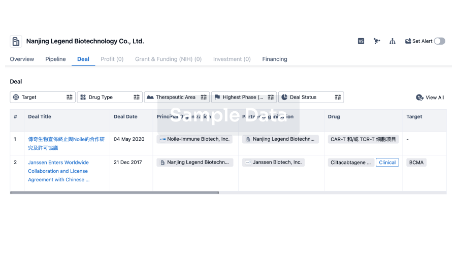
Deal
Boost your decision using our deal data.
login
or
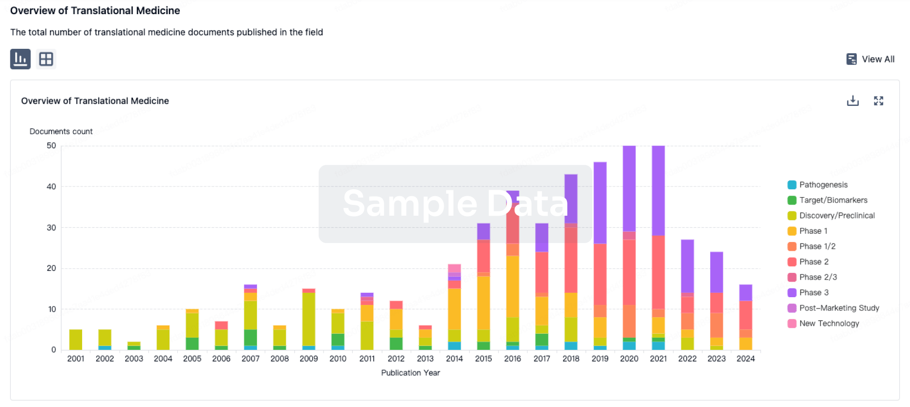
Translational Medicine
Boost your research with our translational medicine data.
login
or
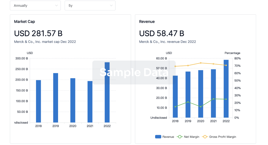
Profit
Explore the financial positions of over 360K organizations with Synapse.
login
or
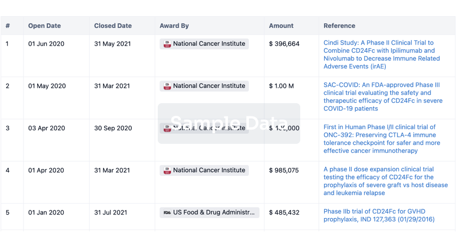
Grant & Funding(NIH)
Access more than 2 million grant and funding information to elevate your research journey.
login
or
Investment
Gain insights on the latest company investments from start-ups to established corporations.
login
or
Financing
Unearth financing trends to validate and advance investment opportunities.
login
or
AI Agents Built for Biopharma Breakthroughs
Accelerate discovery. Empower decisions. Transform outcomes.
Get started for free today!
Accelerate Strategic R&D decision making with Synapse, PatSnap’s AI-powered Connected Innovation Intelligence Platform Built for Life Sciences Professionals.
Start your data trial now!
Synapse data is also accessible to external entities via APIs or data packages. Empower better decisions with the latest in pharmaceutical intelligence.
Bio
Bio Sequences Search & Analysis
Sign up for free
Chemical
Chemical Structures Search & Analysis
Sign up for free