Request Demo
Last update 29 Aug 2025

Gencurix, Inc.
Last update 29 Aug 2025
Overview
Related
1
Clinical Trials associated with Gencurix, Inc.NCT04278469
A Prospective, Randomized, Comparative Study to Evaluate Efficacy of Anticancer Chemotherapy in Predicting Prognosis and Determining Chemotherapy Method in Early Hormone Receptor-positive Breast Cancer Patients With Clinicopathological High Risk and GenesWell™ BCT Low Risk at Multi-center in Korea
A prospective, randomized, comparative study to evaluate efficacy of anticancer chemotherapy in predicting prognosis and determining chemotherapy method in early Hormone Receptor-positive breast cancer patients with clinicopathological high risk and GenesWell™ BCT low risk at multi-center in Korea
Start Date11 Jan 2019 |
Sponsor / Collaborator |
100 Clinical Results associated with Gencurix, Inc.
Login to view more data
0 Patents (Medical) associated with Gencurix, Inc.
Login to view more data
29
Literatures (Medical) associated with Gencurix, Inc.01 Apr 2024·Journal of molecular medicine (Berlin, Germany)
The complement factor H-related protein-5 (CFHR5) exacerbates pathological bone formation in ankylosing spondylitis
Article
Author: Jo, Sungsin ; Lee, Seung Hoon ; Kim, Sang-Hyon ; Han, Jinil ; Youn, Jeehee ; Kim, Tae-Hwan ; Son, Chang-Nam ; Jeon, Chanhyeok ; Kim, Tae-Jong ; Lee, Ji-Hyun ; Kim, Jong-Seo ; Park, Ye-Soo
Ankylosing spondylitis (AS) is a chronic inflammatory disease, characterized by excessive new bone formation. We previously reported that the complement factor H-related protein-5 (CFHR5), a member of the human factor H protein family, is significantly elevated in patients with AS compared to other rheumatic diseases. However, the pathophysiological mechanism underlying new bone formation by CFHR5 is not fully understood. In this study, we revealed that CFHR5 and proinflammatory cytokines (TNF, IL-6, IL-17A, and IL-23) were elevated in the AS group compared to the HC group. Correlation analysis revealed that CFHR5 levels were not significantly associated with proinflammatory cytokines, while CFHR5 levels in AS were only positively correlated with the high CRP group. Notably, treatment with soluble CFHR5 has no effect on clinical arthritis scores and thickness at hind paw in curdlan-injected SKG, but significantly increased the ectopic bone formation at the calcaneus and tibia bones of the ankle as revealed by micro-CT image and quantification. Basal CFHR5 expression was upregulated in AS-osteoprogenitors compared to control cells. Also, treatment with CFHR5 remarkedly induced bone mineralization status of AS-osteoprogenitors during osteogenic differentiation accompanied by MMP13 expression. We provide the first evidence demonstrating that CFHR5 can exacerbate the pathological bone formation of AS. Therapeutic modulation of CFHR5 could be promising for future treatment of AS. KEY MESSAGES: Serum level of CFHR5 is elevated and positively correlated with high CRP group of AS patients. Recombinant CFHR5 protein contributes to pathological bone formation in in vivo model of AS. CFHR5 is highly expressed in AS-osteoprogenitors compared to disease control. Recombinant CFHR5 protein increased bone mineralization accompanied by MMP13 in vitro model of AS.
01 Apr 2022·Laboratory investigation; a journal of technical methods and pathologyQ2 · MEDICINE
Matrix metalloproteinase 11 (MMP11) in macrophages promotes the migration of HER2-positive breast cancer cells and monocyte recruitment through CCL2–CCR2 signaling
Q2 · MEDICINE
Article
Author: Han, Jinil ; Shin, Young Kee ; Kwon, Mi Jeong ; Jeong, Hyojin ; Yang, Hobin ; Chae, Ha Yeong ; Sung, Chang Ohk ; Kang, Shin Ung ; Cho, Soo Youn ; Choi, Yoon-La
Matrix metalloproteinase 11 (MMP11), a member of the MMP family involved in the degradation of the extracellular matrix, has been implicated in cancer progression. Despite the stromal expression of MMP11 in breast cancer, the prognostic significance and role of MMP11 in immune or stromal cells of breast cancer remain unclear. Based on the immunohistochemical analysis of breast cancer tissues from 497 patients, we demonstrated that MMP11 expression in mononuclear inflammatory cells (predominantly macrophages) is an independent negative prognostic factor in breast cancer, whereas MMP11 expression in tumor cells and fibroblasts is not associated with patient survival. Enforced MMP11 expression in breast cancer cells did not promote cell proliferation and migration. However, MMP11-overexpressing macrophages enhanced the migration of HER2-positive (HER2+) breast cancer cells, recruitment of monocytes, and tube formation of endothelial cells. Furthermore, we found that the chemokine CCL2 secreted from MMP11-overexpressing macrophages activated the MAPK pathway via its receptor CCR2 in breast cancer cells, thereby promoting the migration of HER2+ breast cancer cells through MMP9 upregulation. We also found that MMP11 expression in macrophages was stimulated by MMP11-overepressing HER2+ breast cancer cells. Collectively, our findings provide evidence that MMP11 in macrophages may play a pro-tumoral role in HER2+ breast cancer through interaction with cancer cells, monocytes, and endothelial cells.
01 Jan 2020·Cellular and molecular neurobiology
Bcl-2 Overexpression Induces Neurite Outgrowth via the Bmp4/Tbx3/NeuroD1 Cascade in H19-7 Cells
Article
Author: Han, Joong-Soo ; Lee, So Young ; Park, Shin-Young ; Han, Jinil ; Kang, Min-Jeong ; Lee, Yun Young ; Choi, Hye-Jin
Bcl-2 is overexpressed in the nervous system during neural development and plays an important role in modulating cell survival. In addition to its anti-apoptotic function, it has been suggested previously that Bcl-2 might act as a mediator of neuronal differentiation. However, the mechanism by which Bcl-2 might influence neurogenesis is not sufficiently understood. In this study, we aimed to determine the non-apoptotic functions of Bcl-2 during neuronal differentiation. First, we used microarrays to analyze the whole-genome expression patterns of rat neural stem cells overexpressing Bcl-2 and found that Bcl-2 overexpression induced the expression of various neurogenic genes. Moreover, Bcl-2 overexpression increased the neurite length as well as expression of Bmp4, Tbx3, and proneural basic helix-loop-helix genes, such as NeuroD1, NeuroD2, and Mash1, in H19-7 rat hippocampal precursor cells. To determine the hierarchy of these molecules, we selectively depleted Bmp4, Tbx3, and NeuroD1 in Bcl-2-overexpressing cells. Bmp4 depletion suppressed the upregulation of Tbx3 and NeuroD1 as well as neurite outgrowth, which was induced by Bcl-2 overexpression. Although Tbx3 knockdown repressed Bcl-2-mediated neurite elaboration and downregulated NeuroD1 expression, it did not affect Bcl-2-induced Bmp4 expression. While the depletion of NeuroD1 had no effect on the expression of Bcl-2, Bmp4, or Tbx3, Bcl-2-mediated neurite outgrowth was suppressed. Taken together, these results demonstrate that Bcl-2 regulates neurite outgrowth through the Bmp4/Tbx3/NeuroD1 cascade in H19-7 cells, indicating that Bcl-2 may have a direct role in neuronal development in addition to its well-known anti-apoptotic function in response to environmental insults.
5
News (Medical) associated with Gencurix, Inc.19 Jun 2025
GENCURIX becomes inaugural partner in QIAGEN’s QIAcuityDx™ Partnering Program to drive global-standard precision diagnostics
SEOUL, SOUTH KOREA, June 19, 2025 /
EINPresswire.com
/ -- GENCURIX, Inc., a leading molecular diagnostics company specializing in oncology, today announced that it has signed a global development and commercialization agreement with QIAGEN, a global leader in molecular diagnostics, to jointly develop and commercialize digital PCR-based oncology diagnostic products.
Under the agreement, GENCURIX will develop in vitro diagnostic (IVD) kits utilizing QIAGEN’s digital PCR system, QIAcuityDx Four, while QIAGEN will hold exclusive global distribution rights. The products will be co-branded under QIAGEN Partner
®
as well as both companies’ brand names.
Importantly, GENCURIX becomes the first official development partner in QIAGEN’s newly launched QIAcuityDx Partnering Program, which aims to create a robust menu of clinical diagnostic assays on the QIAcuityDx Four platform. This positions GENCURIX at the forefront of a new global initiative to accelerate precision diagnostics in oncology.
Through this collaboration, GENCURIX will integrate its proprietary molecular diagnostic technologies and biomarker-driven cancer detection capabilities with QIAGEN’s digital PCR and software technologies to develop IVD kits for various cancer types. These kits will be marketed globally through QIAGEN’s established commercial infrastructure, ensuring broad and streamlined access for clinical laboratories.
The partnership will begin with a stepwise development of multiple oncology-focused IVD products, jointly pre-agreed by both parties.
“This strategic partnership with QIAGEN represents a major inflection point for expanding our oncology molecular diagnostic technologies into the global market,” said Sang Rae Cho, CEO of GENCURIX. “We are confident that the synergy between our advanced diagnostic content and QIAGEN’s high-performance platform will lead to global-standard precision cancer diagnostic solutions.”
“The QIAcuityDx Partnering Program is designed to enable the generation of a broad menu of IVD assays on the platform,” said Jonathan Arnold, Vice President and Head, Partnering for Precision Diagnostics at QIAGEN. “Our first partnership with GENCURIX is an exciting launch that gives oncology labs access to high-quality IVD assays that complement qPCR and NGS. We look forward to working with GENCURIX to grow this important program.”
With this agreement, GENCURIX strengthens its position as a global innovator in multiplex assay development, especially for tissue and liquid biopsy applications. The partnership also introduces scalable commercialization opportunities, positioning GENCURIX to rapidly bring innovative cancer diagnostics to global markets through QIAGEN’s extensive sales and technical support network.
Gencurix
Gencurix
email us here
Legal Disclaimer:
EIN Presswire provides this news content "as is" without warranty of any kind. We do not accept any responsibility or liability
for the accuracy, content, images, videos, licenses, completeness, legality, or reliability of the information contained in this
article. If you have any complaints or copyright issues related to this article, kindly contact the author above.
Diagnostic Reagents
16 Jan 2024
DUBLIN--(
BUSINESS WIRE
)--The
"Acquired Gene or Chromosome Alterations Pipeline Report including Stages of Development, Segments, Region and Countries, Regulatory Path and Key Companies, 2023 Update"
report has been added to
ResearchAndMarkets.com's
offering.
Acquired Gene or Chromosome Alterations Pipeline Report provides comprehensive information about the Acquired Gene or Chromosome Alterations pipeline products with comparative analysis of the products at various stages of development and information about the clinical trials which are in progress.
This report offers extensive coverage of Acquired Gene or Chromosome Alterations under development, providing comprehensive insights into pipeline products, major players, developmental activities, clinical trial data, and recent developments in the segment/industry.
Acquired Gene or Chromosome Alterations comprises of reagents employed for genetic testing to detect cancer causing/ related alterations, such as; k-ras, BRCA 1+2, Telomerase and HER-2/neu (human epidermal growth factor receptor 2).
The report reviews the details of major pipeline products, including product descriptions, licensing and collaboration details, and other developmental activities related to Acquired Gene or Chromosome Alterations. It also lists all the pipeline projects involving major players engaged in the development of Acquired Gene or Chromosome Alterations.
Coverage of pipeline products is based on various stages of development, ranging from Early Development to Approved/Issued stages, allowing readers to understand the progress of these products in the pipeline. Key clinical trial data related to ongoing trials specific to pipeline products in the Acquired Gene or Chromosome Alterations segment is provided, offering valuable insights into the clinical development of these products.
Reasons to Buy
Formulate significant competitor information, analysis, and insights to improve R&D strategies
Identify emerging players with potentially strong product portfolio and create effective counter strategies to gain competitive advantage
Identify and understand important and diverse types of Acquired Gene or Chromosome Alterations under development
Develop market-entry and market expansion strategies
Plan mergers and acquisitions effectively by identifying major players with the most promising pipeline
In-depth analysis of the product's current stage of development, territory and estimated launch date
Key Topics Covered:
1 Table of Contents
1.1 List of Tables
1.2 List of Figures
2 Introduction
2.1 Acquired Gene or Chromosome Alterations Overview
3 Products under Development
3.1 Acquired Gene or Chromosome Alterations - Pipeline Products by Stage of Development
3.2 Acquired Gene or Chromosome Alterations - Pipeline Products by Territory
3.3 Acquired Gene or Chromosome Alterations - Pipeline Products by Regulatory Path
3.4 Acquired Gene or Chromosome Alterations - Pipeline Products by Estimated Approval Date
3.5 Acquired Gene or Chromosome Alterations - Ongoing Clinical Trials
4 Acquired Gene or Chromosome Alterations - Pipeline Products under Development by Companies
4.1 Acquired Gene or Chromosome Alterations Companies - Pipeline Products by Stage of Development
4.2 Acquired Gene or Chromosome Alterations - Pipeline Products by Stage of Development
5 Acquired Gene or Chromosome Alterations Companies and Product Overview
6 Acquired Gene or Chromosome Alterations- Recent Developments
7 Appendix
A selection of companies mentioned in this report includes
Abital Pharma Pipelines Ltd
Admera Health LLC
Agena Bioscience Inc
Aix-Marseille University
Albert Ludwigs University of Freiburg
Apteryx Imaging Inc
Baruch S. Blumberg Institute
Baylor College of Medicine
bioMerieux SA
Biotype Innovation GmbH
Blondin Bioscience, LLC
Cardiff Oncology Inc
Cepheid Inc
Children's Hospital Los Angeles
Children's Hospital of Philadelphia
China Medical Technologies Inc (Inactive)
Chronix Biomedical Inc
Columbia University
Duke University
Empire Genomics LLC
Fred Hutchinson Cancer Research Center
French National Institute of Health and Medical Research
Garvan Institute of Medical Research
Gencurix Inc
Genetag Technology, Inc
Genomic Health Inc
GenomicTree Co Ltd
German Cancer Research Center
H. Lee Moffitt Cancer Center & Research Institute Inc
Heali Ltd
Hebrew University of Jerusalem
Heidelberg Pharma AG
Helmholtz Centre for Infection Research
Inform Genomics Inc
Institute of Cancer Research
Interpace Biosciences Inc
iTP Biomedica Corp
Johns Hopkins University
Massachusetts General Hospital
Mayo Clinic
McGill University
MDNA Life Sciences Inc
MolecularMD Corp
Myriad International GmbH
Nel ASA
Northern California Institute for Research and Education
NucleoBio Inc
Ohio State University
OmicsWay Corp
Oxford Biodynamics Plc
Oxford Cancer Biomarkers Ltd
Pennsylvania State University
Princess Margaret Cancer Centre
QuantuMDx Group Ltd
Queen's University Belfast
Rosetta Genomics Ltd (Inactive)
Stanford University
Stony Brook University
Syamala Srinivasa Life Sciences Pvt. Ltd
Telo Genomics Corp
The Chinese University of Hong Kong
The University of New South Wales press Limited
The Walter and Eliza Hall Institute of Medical Research
TheraDiag SA
Therawis Diagnostics GmbH
Thrive Earlier Detection Corp
Total Brain Ltd
Trinity College Dublin
University College London
University of California San Diego
University of Cambridge
University of Central Florida
University of Cologne
University of Copenhagen
University of Hawaii at Manoa
University of Montreal
University of Rochester
University of South Carolina
University of Southern California
University of Strathclyde
University of Sydney
University of Texas Medical Branch at Galveston
University of Washington
Vyant Bio Inc
Wistar Institute
Yaathum Biotech Pvt Ltd
Yale University
For more information about this report visit
https://www.researchandmarkets.com/r/fb39kc
About ResearchAndMarkets.com
ResearchAndMarkets.com is the world's leading source for international market research reports and market data. We provide you with the latest data on international and regional markets, key industries, the top companies, new products and the latest trends.
Acquisition
14 Jun 2023
SEOUL, South Korea, June 13, 2023 /PRNewswire/ -- Gencurix, a leading cancer molecular diagnostics company, has obtained approval from the Korean Ministry of Food and Drug Safety for its Droplex EGFR Mutation Test v2. This product is a companion diagnostic test designed to detect EGFR mutations commonly found in NSCLC patients and guide the selection of appropriate targeted anticancer therapies based on the test results. It is an IVD product that can be used with Bio-Rad's Droplet Digital PCR instrument and has also obtained CE certification in Europe last year.
Compared to existing EGFR mutation tests using RT-PCR, this product demonstrates significantly higher sensitivity. This makes it capable of accurately detecting mutations even when plasma samples are used as the specimen. Additionally, while most existing products detect fewer than 50 mutations, this test can detect up to 107 mutations, further enhancing its accuracy.
Notably, this product has an additional strength in its ability to detect Exon 20 Insertion, a mutation that has recently garnered significant attention in the oncologist community following the launches of Janssen's Rybrevant and Takeda's Exkivity. Among the existing single gene tests, there were no tests available to properly detect this mutation, necessitating the use of costly and time-consuming NGS tests.
This is the second EGFR mutation test for which Gencurix has obtained approval. In addition, Gencurix has obtained European CE registration for a total of nine products, including KRAS, BRAF, C-MET, PIK3CA, ESR1, and POLE tests, making it the company with the highest number of digital PCR-based IVD tests in its portfolio.
Hyun Park, VP of Strategy & Business Development at Gencurix, stated, "Gencurix has been developing diagnostic products that utilize Droplet Digital PCR technology, which has primarily been used for research purposes, in the field of cancer diagnostics. Through years of research and development, we are proud to have achieved world-class technological capabilities and a product portfolio." Gencurix is also developing solutions for early cancer detection and minimal residual disease (MRD) screening using digital PCR technology. The company also introduced new technology detecting colorectal cancer recurrence at AACR last April.
Gencurix has been supplying diagnostic kits to major hospitals in Korea, such as Samsung Medical Center and Yonsei Severance Hospital, and has been actively expanding its global sales network, securing distributorship agreements in Europe, Asia, and other continents. In addition to collaborating with Bio-Rad, a leading manufacturer of digital PCR instruments, on marketing efforts, Gencurix is also pursuing the acquisition of new distributors.
About Gencurix:
Gencurix is a leading provider of molecular cancer diagnostics, dedicated to developing innovative solutions that improve patient outcomes. Committed to research and development, Gencurix strives to address unmet needs in cancer diagnosis and treatment, ultimately revolutionizing the field of precision medicine. For more information, visit .
SOURCE Gencurix
AcquisitionDiagnostic Reagents
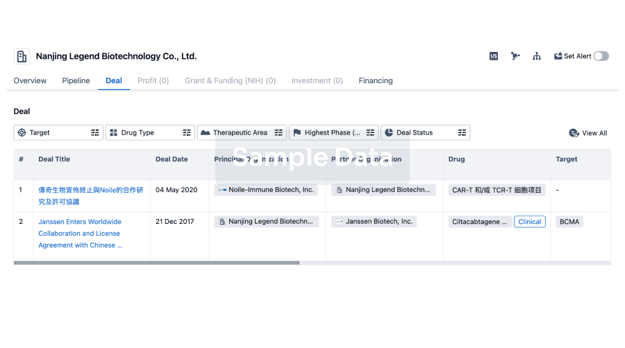
100 Deals associated with Gencurix, Inc.
Login to view more data
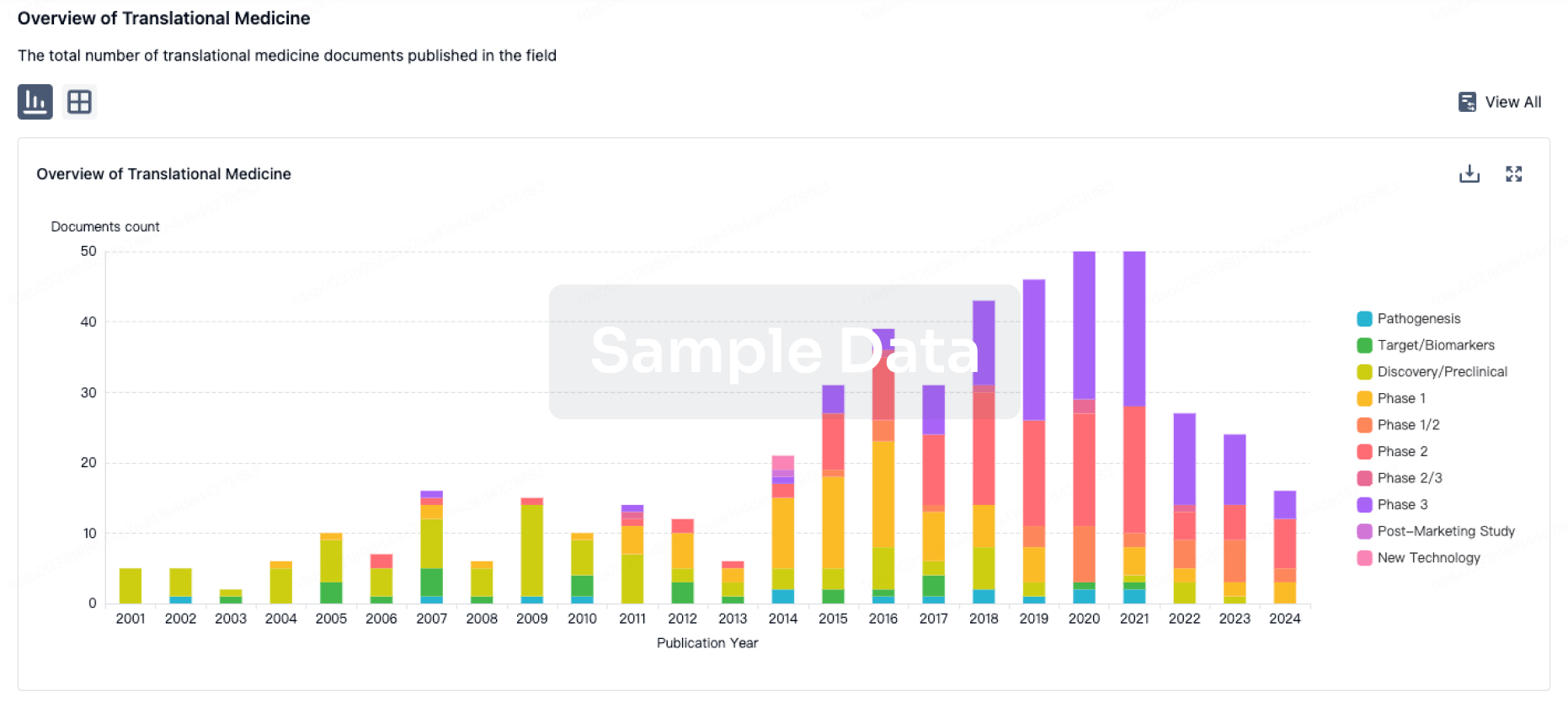
100 Translational Medicine associated with Gencurix, Inc.
Login to view more data
Corporation Tree
Boost your research with our corporation tree data.
login
or

Pipeline
Pipeline Snapshot as of 13 Dec 2025
No data posted
Login to keep update
Deal
Boost your decision using our deal data.
login
or

Translational Medicine
Boost your research with our translational medicine data.
login
or

Profit
Explore the financial positions of over 360K organizations with Synapse.
login
or

Grant & Funding(NIH)
Access more than 2 million grant and funding information to elevate your research journey.
login
or

Investment
Gain insights on the latest company investments from start-ups to established corporations.
login
or

Financing
Unearth financing trends to validate and advance investment opportunities.
login
or

AI Agents Built for Biopharma Breakthroughs
Accelerate discovery. Empower decisions. Transform outcomes.
Get started for free today!
Accelerate Strategic R&D decision making with Synapse, PatSnap’s AI-powered Connected Innovation Intelligence Platform Built for Life Sciences Professionals.
Start your data trial now!
Synapse data is also accessible to external entities via APIs or data packages. Empower better decisions with the latest in pharmaceutical intelligence.
Bio
Bio Sequences Search & Analysis
Sign up for free
Chemical
Chemical Structures Search & Analysis
Sign up for free