Request Demo
What is the mechanism of Ferucarbotran?
17 July 2024
Ferucarbotran is a superparamagnetic iron oxide nanoparticle (SPION) commonly used as a contrast agent in magnetic resonance imaging (MRI). This agent is particularly valuable in enhancing the visibility of internal body structures, making it easier to diagnose and monitor various medical conditions. To understand the mechanism of Ferucarbotran, it is essential to delve into its composition, properties, and how it interacts with the body during imaging procedures.
Ferucarbotran is composed of iron oxide cores coated with a carbohydrate layer, which stabilizes the particles and facilitates their dispersion in aqueous solutions. The superparamagnetic nature of these nanoparticles arises from their iron oxide cores, typically made of magnetite (Fe3O4) or maghemite (γ-Fe2O3). These cores are small enough, usually in the range of 10-50 nanometers, to exhibit superparamagnetic behavior. This means that in the presence of an external magnetic field, the nanoparticles can become magnetized but will lose their magnetization once the field is removed. This property is crucial for their function in MRI.
When Ferucarbotran is injected into the body, it circulates through the bloodstream and is eventually taken up by the reticuloendothelial system (RES), which includes the liver, spleen, and lymph nodes. The carbohydrate coating on the nanoparticles ensures biocompatibility and minimizes any potential toxicity. The uptake by the RES is primarily due to the recognition of these particles by macrophages, which are part of the body's immune system.
In the context of MRI, Ferucarbotran serves as a negative contrast agent, primarily affecting the T2 relaxation time of protons in water molecules within tissues. The superparamagnetic properties of the iron oxide cores create local magnetic field inhomogeneities when exposed to the external magnetic field of the MRI machine. These inhomogeneities lead to rapid dephasing of proton spins in the surrounding water molecules, resulting in a significant reduction in T2 relaxation time. Consequently, tissues containing Ferucarbotran appear darker on T2-weighted MRI images.
The primary application of Ferucarbotran in MRI is for liver imaging. The liver's rich macrophage population avidly takes up these nanoparticles, allowing for enhanced contrast between healthy liver tissue and abnormal lesions such as tumors or fibrosis. Tumorous tissues, which typically have fewer macrophages, do not take up Ferucarbotran as efficiently, making them appear as hyperintense (brighter) areas against the darkened background of normal liver tissue on T2-weighted images. This contrast enhancement greatly aids in the detection and characterization of liver lesions.
Beyond liver imaging, Ferucarbotran can also be used to assess lymph nodes and spleen pathology due to its uptake by the RES. Its application extends to other areas where the visualization of macrophage activity or the RES can provide valuable diagnostic information.
In summary, the mechanism of Ferucarbotran revolves around its superparamagnetic iron oxide cores and carbohydrate coating, which facilitate its stability, biocompatibility, and uptake by the reticuloendothelial system. Upon administration, these nanoparticles enhance the contrast in MRI images by significantly reducing the T2 relaxation time of surrounding water protons, leading to darker images of tissues that have absorbed the agent. This property is particularly useful in liver imaging and other RES-related diagnostic applications. Understanding this mechanism helps in appreciating the role of Ferucarbotran in improving the accuracy and effectiveness of MRI diagnostics.
How to obtain the latest development progress of all drugs?
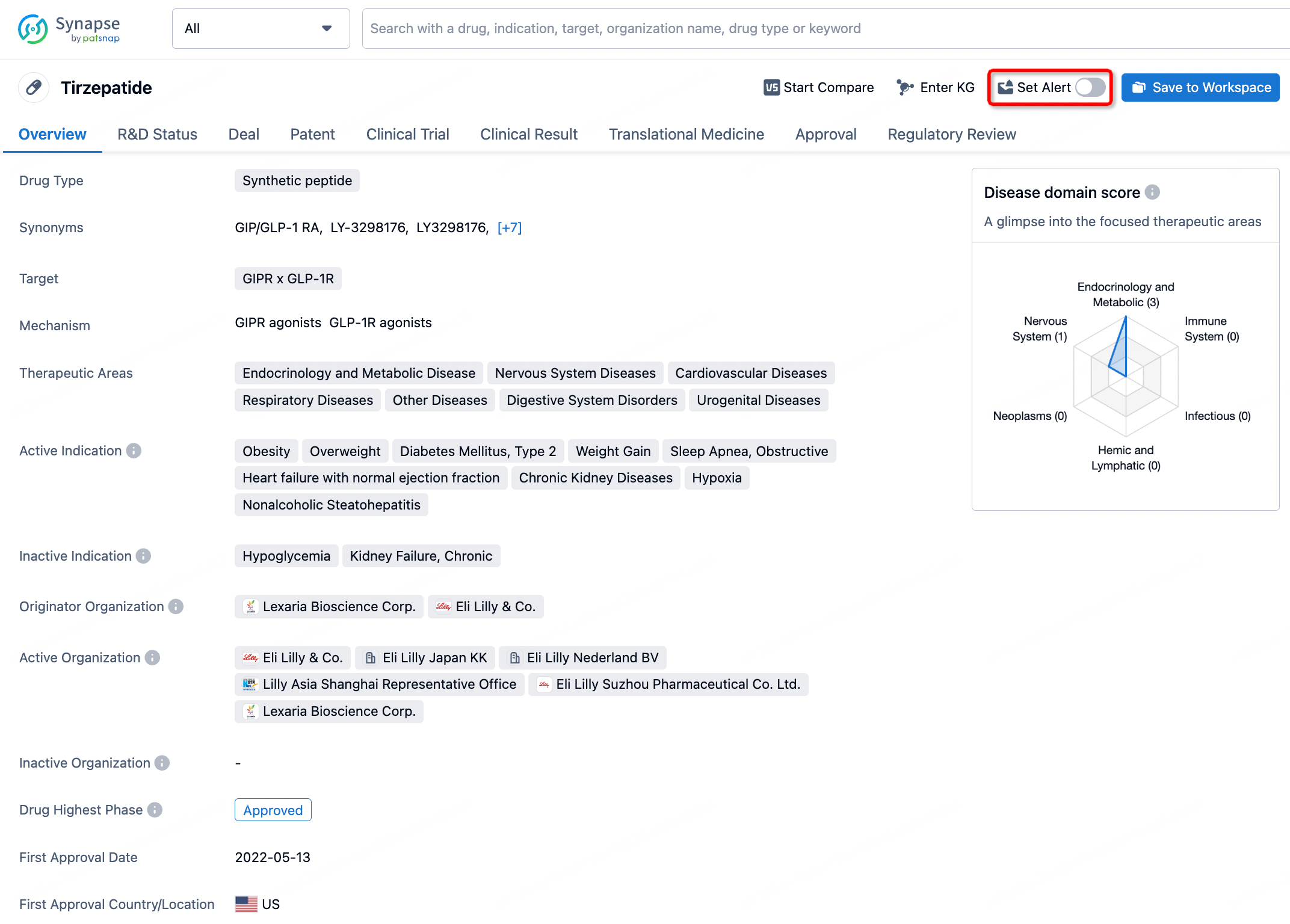
In the Synapse database, you can stay updated on the latest research and development advances of all drugs. This service is accessible anytime and anywhere, with updates available daily or weekly. Use the "Set Alert" function to stay informed. Click on the image below to embark on a brand new journey of drug discovery!


AI Agents Built for Biopharma Breakthroughs
Accelerate discovery. Empower decisions. Transform outcomes.
Get started for free today!
Accelerate Strategic R&D decision making with Synapse, PatSnap’s AI-powered Connected Innovation Intelligence Platform Built for Life Sciences Professionals.
Start your data trial now!
Synapse data is also accessible to external entities via APIs or data packages. Empower better decisions with the latest in pharmaceutical intelligence.