Request Demo
What is the mechanism of Iohexol?
17 July 2024
Iohexol is a nonionic, water-soluble contrast agent widely used in medical imaging, particularly in procedures such as computed tomography (CT) scans, angiography, and myelography. Understanding the mechanism of Iohexol involves delving into its chemical structure, pharmacokinetics, and the way it interacts with biological tissues to enhance imaging.
Chemically, Iohexol belongs to the class of low-osmolar contrast agents (LOCAs). Its molecular structure is based on an iodinated benzene ring, which is crucial for its radiopacity. The high atomic number of iodine (Z=53) makes it an excellent agent for absorbing X-rays and producing contrast. This iodinated benzene ring is substituted with several hydrophilic groups, such as hydroxyl and carboxamide groups, which significantly reduce its osmolality compared to older high-osmolar contrast media (HOCM). The lower osmolality translates to fewer adverse reactions, making Iohexol safer for patient use.
Once administered, typically via intravenous injection, Iohexol rapidly distributes through the extracellular fluid. Its hydrophilic nature prevents significant crossing of cellular membranes, thus confining it mainly to extracellular spaces. This characteristic is particularly advantageous in imaging, as it allows for clear delineation of vascular structures and interstitial spaces.
The pharmacokinetics of Iohexol are relatively straightforward. After administration, it disperses quickly and is not metabolized by the body. It remains relatively unchanged and is excreted primarily by the kidneys via glomerular filtration. The half-life of Iohexol in individuals with normal renal function is approximately 2 hours. However, in patients with renal impairment, the excretion can be significantly delayed, necessitating careful monitoring and possible dosage adjustments.
The mechanism of action of Iohexol in imaging is primarily based on its ability to enhance the contrast of structures within the body under X-ray examination. The iodine atoms absorb X-rays more effectively than surrounding tissues, creating a clear distinction on radiographic images. This heightened contrast allows radiologists to obtain detailed images of blood vessels, organs, and other tissues, facilitating accurate diagnosis and evaluation.
In vascular imaging, for instance, Iohexol is injected into the bloodstream and highlights blood vessels, enabling the visualization of blockages, aneurysms, or other vascular abnormalities. In CT imaging, Iohexol distributes into the extracellular space, enhancing the contrast between different tissue types, which aids in the identification of tumors, infections, or other pathological conditions.
While Iohexol is generally well-tolerated, it is not devoid of side effects. Some patients may experience mild reactions such as nausea, vomiting, or a warm sensation during injection. More severe reactions, although rare, can include allergic responses, renal impairment, or cardiovascular events. Therefore, patient screening and proper hydration are essential to minimize risks.
In conclusion, Iohexol serves as a crucial tool in medical imaging due to its efficient contrast-enhancing properties, favorable safety profile, and predictable pharmacokinetics. Its mechanism, rooted in its iodinated structure and hydrophilic nature, allows for clear and detailed imaging of various body structures, significantly aiding in diagnostic accuracy and patient care. Understanding this mechanism is vital for healthcare professionals to optimize its use and ensure patient safety during imaging procedures.
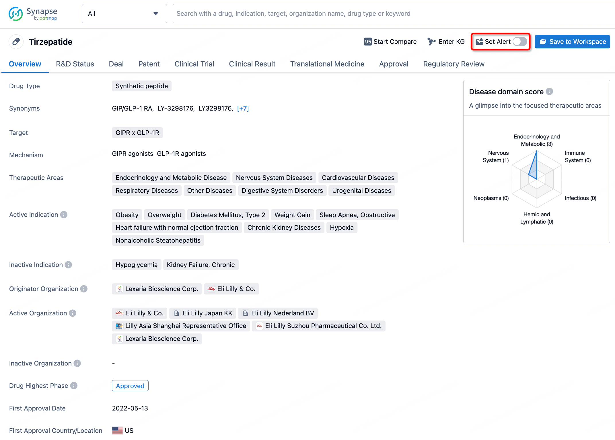
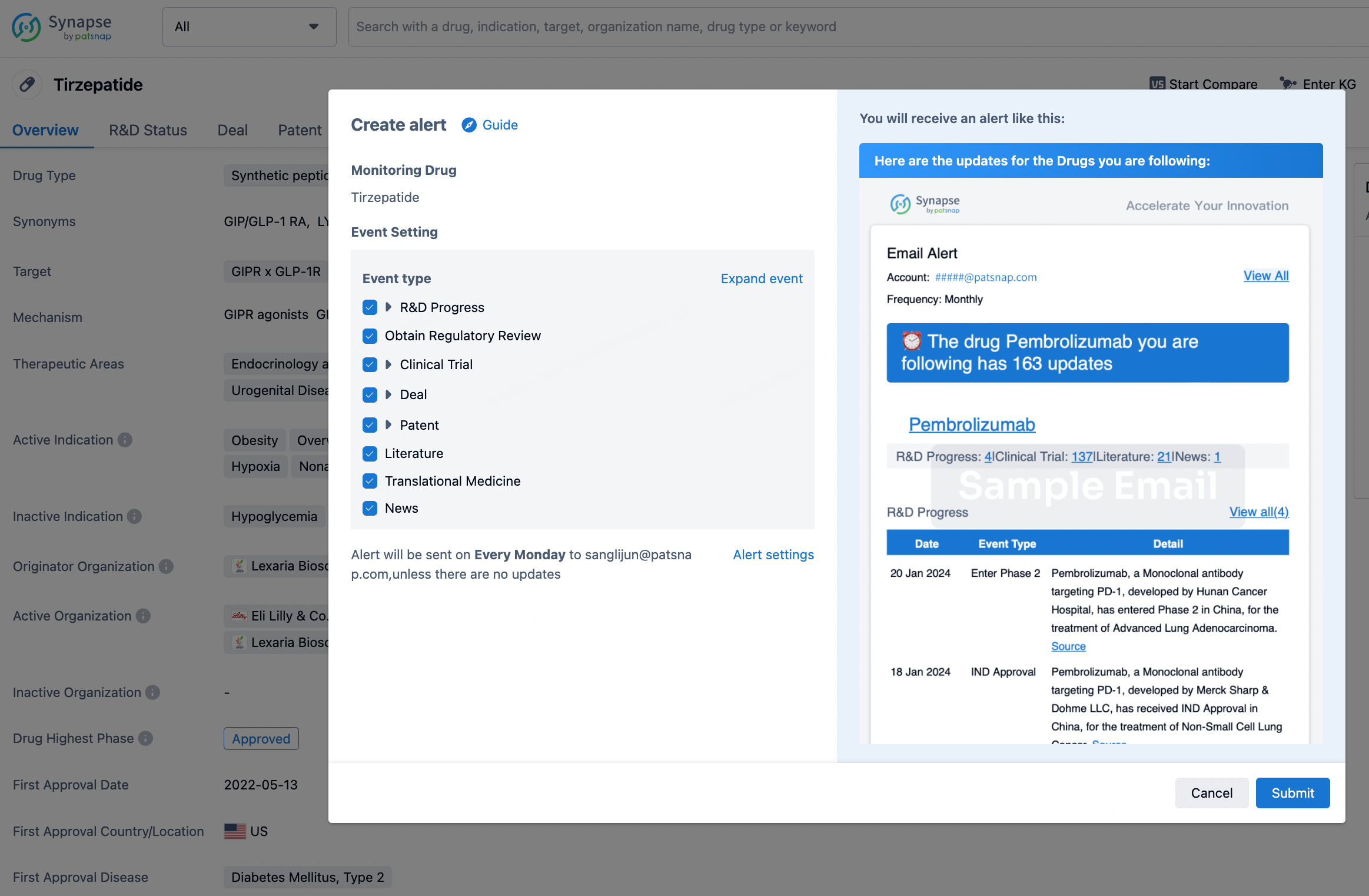
How to obtain the latest development progress of all drugs?
In the Synapse database, you can stay updated on the latest research and development advances of all drugs. This service is accessible anytime and anywhere, with updates available daily or weekly. Use the "Set Alert" function to stay informed. Click on the image below to embark on a brand new journey of drug discovery!

AI Agents Built for Biopharma Breakthroughs
Accelerate discovery. Empower decisions. Transform outcomes.
Get started for free today!
Accelerate Strategic R&D decision making with Synapse, PatSnap’s AI-powered Connected Innovation Intelligence Platform Built for Life Sciences Professionals.
Start your data trial now!
Synapse data is also accessible to external entities via APIs or data packages. Empower better decisions with the latest in pharmaceutical intelligence.