Request Demo
Last update 11 Jul 2026
Zanolimumab
Last update 11 Jul 2026
Overview
Basic Info
Drug Type Monoclonal antibody |
Synonyms Anti-CD4 monoclonal antibody MDX CD4, Zanolimumab (USAN/INN), HUMAX-CD4 + [4] |
Target |
Action inhibitors |
Mechanism CD4 inhibitors(T-cell surface antigen CD4 inhibitors) |
Therapeutic Areas |
Active Indication- |
Inactive Indication |
Originator Organization |
Active Organization- |
Inactive Organization |
License Organization |
Drug Highest PhaseDiscontinuedPhase 3 |
First Approval Date- |
RegulationOrphan Drug (European Union) |


Login to view timeline
Structure/Sequence
Sequence Code 28264011H
Sequence Code 28264012L
Related
7
Clinical Trials associated with ZanolimumabNCT01160445
Phase II Study of Aldesleukin (IL-2) Following the Administration of Zanolimumab (Anti-CD4mAb) in Metastatic Melanoma and Metastatic Renal Cancer

NCT00893516
A Multicentre Open-label Dose Escalation Tiral of Zanolimumab in Combination With CHOP Chemotherapy in Patients With CD4 Positive Non-cutaneous Peripheral T-cell Lymphoma With Nodal Involvement.
NCT00877656
An Open-Label Therapeutic Exploratory Clinical Trial of HuMax-CD4, a Fully Human Monoclonal Anti-CD4 Antibody, in Patients With Refractory or Relapsed Non-Cutaneous CD4+ T-Cell Lymphoma
100 Clinical Results associated with Zanolimumab
Login to view more data
100 Translational Medicine associated with Zanolimumab
Login to view more data
100 Patents (Medical) associated with Zanolimumab
Login to view more data
21
Literatures (Medical) associated with Zanolimumab01 Aug 2011British journal of haematology
Alemtuzumab‐resistant Sézary syndrome responding to zanolimumab*
Letter
Author: Martin J. S. Dyer ; W. Mark Bamford ; Volha Shpadaruk ; D. Ben J. Kennedy ; Anton B. Alexandroff ; Robert Burd
Primary cutaneous T-cell lymphomas (CTCL) are rare malignancies characterized by clonal T-cell infiltrates (Kim et al, 2005; Prince et al, 2009). Mycosis fungoides (MF), the most common form of CTCL, presents with patches, plaques, tumours and erythroderma whereas Sézary syndrome (SS) is characterized by erythroderma, lymphadenopathy, and Sézary cells in the peripheral blood. Although CTCL often pursues an indolent course, a poor prognosis has been reported in patients with tumoural/ulcerated lesions, erythroderma, and transformation into T-cell lymphoma (Kim et al, 2005; Prince et al, 2009). Advanced stages of CTCL often show resistance to available treatment (Kim et al, 2005; Cheson, 2007; Prince et al, 2009). Two unconjugated humanized monoclonal antibodies (MAbs) have been used recently in the treatment of CTCL. Alemtuzumab, a CD52 MAb, has proved a valuable treatment option for several subtypes of T-cell malignancy. In CTCL, clinical response rates of 84% (47% complete remission, CR) have been demonstrated in SS patients (Ravandi & O’Brien, 2005; Bernengo et al, 2007). Long-term, durable responses may be obtained with alemtuzumab (DBJK and MJSD, unpublished observations). Zanolimumab is a CD4 MAb that has shown activity in patients with CTCL and non-CTCL lymphomas (d’Amore et al, 2010). In two Phase II studies, 56% of MF patients responded to high dose of zanolimumab whilst for SS patients the response rates were 25% (Kim et al, 2007; d’Amore et al, 2010). Here, we present a case of SS who, whilst initially sensitive to alemtuzumab, became alemtuzumab-resistant due to selective loss of CD52 expression. Nevertheless, it was possible to induce a second durable remission with zanolimumab. A 68-year-old Caucasian woman was diagnosed with SS in 2001. She presented with ‘resistant-to-treatment eczema’ and inconclusive skin biopsies, but later developed extensive exfoliative erythema. There was a low-level lymphocytosis composed of Sézary cells expressing CD4 and CD52. Polymerase chain reaction of the T-cell receptor γ locus (TRG@) of skin, lymph node and peripheral blood samples showed identical clonal rearrangements. Full body computerized tomography (CT) scan was normal. The patient was managed with topical and systemic steroids, PUVA (psoralen and ultraviolet A) photochemotherapy, ciclosporin, methotrexate and acitretin with little effect. By 2004, she had developed erythroderma and was symptomatic with fever, sweats and shivering (Fig 1A). Skin biopsy showed a mild upper dermal inflammatory infiltrate composed of small lymphocytes with extension into the overlying epidermis with associated spongiosis. No classical Pautrier microabscesses were observed. Malignant T-cells expressed CD4 (Fig 1B). She was treated with subcutaneous alemtuzumab 30 mg initially on a daily dose schedule. Her symptoms resolved within 72 h. After the first week, doses of alemtuzumab were given three times per week for a total of 12 weeks. This therapy induced a CR that lasted for 18 months. (A) Generalized, exfoliative erythroderma seen in this patient before treatment with either alemtuzumab or zanolimumab. (B) Skin biopsy stained with CD4 showing a mild upper dermal infiltrate composed of generally small lymphocytes extending into the epidermis with associated spongiosis. No classical Pautrier microabscesses were observed (Original magnification ×20). At relapse, there were no Sézary cells in the peripheral blood. Alemtuzumab was reinstated but neither this nor intravenously administered MAb nor the addition of high dose methylprednisolone made any significant impression on her disease. Neutralizing alemtuzumab anti-idiotype antibodies were sought but none detected. Alemtuzumab was abandoned. Two months later the patient was commenced on zanolimumab. Zanolimumab (980 mg per infusion) was administered initially twice a week for 2 weeks. The patient’s symptoms and erythroderma were slow to resolve but after 4 months of treatment (17 infusions), she had achieved almost CR: the patient was well and had only mild erythema on thighs and torso. Analysis of peripheral blood identified no detectable CD4 lymphocytes. Zanolimumab was discontinued and CD4 lymphopenia resolved slowly over a period of 12 months without any infective complications. One month after stopping zanolimumab the patient suddenly developed macular, blanching, urticarial patches affecting 80% of body and became systemically unwell (Fig 2). Investigations showed normal C-reactive protein and full blood count. Skin cultures yielded Staphylococcus aureus and beta-hemolytic Streptococcus group B. Skin biopsy exhibited non-specific findings and showed no histopathological, immunocytochemical or molecular features of CTCL. The patient received erythromycin and mupirocin with resolution of signs and symptoms within 2 weeks. She remained well until she relapsed with erythroderma and peripheral blood Sézary cells 12 months later. At the time of relapse, peripheral blood Sézary cells expressed CD4 but not CD52, suggesting that the lack of efficacy of alemtuzumab on the second treatment was due to selection of cells lacking expression of CD52. Labile urticarial eruption affecting 80% of the body surface following zanolimumab. Zanolimumab blocks T-cell activation via CD4 and also facilitates destruction of target cells via antibody-dependent cellular cytotoxicity (ADCC) (Rider et al, 2007); which of these two possible mechanisms is of pivotal importance in the treatment of T-cell malignancy is not yet clear. In comparison with alemtuzumab, where therapeutic responses are (as in our patient) often very rapid, responses to zanolimumab are usually slow with a median time to response of between 2 and 12 weeks) (Kim et al, 2007). These data may indicate that the major mode of action of this MAb is through blocking of T-cell activation rather than ADCC. This is supported by the observation that CD4 internalises after zanolimumab cross-linking, rendering cells effectively CD4 negative. In contrast, alemtuzumab does not internalize significantly and remains available to mediate ADCC even in vivo (Dyer et al, 1989). It is interesting to note that in this case the CD4 expression was maintained after zanolimumab therapy, whilst CD52 expression was undetectable. The optimal doses of zanolimumab, the role of zanolimumab in combination with other agents and, in particular, the possible role of maintenance zanolimumab therapy in responding patients remains to be determined. Despite the complete absence of CD4 cells in the peripheral blood that follows its use, the MAb does not appear to be associated with a significant increase in opportunistic infections, which may allow for more protracted courses of this MAb to be given in order to improve and maintain responses. Our patient experienced an apparent ‘flare’ of disease but this was due to Staphylococcus aureus infection. Exacerbation of CTCL triggered by Staphylococcus aureus has been reported previously. It should also be noted that eczematous dermatitis has also been observed in a proportion of patients receiving zanolimumab and appears to correlate with response. We thank Genmab (Copenhagen DK) for generously providing zanolimumab for our patient on compassionate request basis; Genmab has licensed zanolimumab to TenX Biopharma Inc (Philadelphia, PA) for further development and commercialization. We thank Professor Geoff Hale (BioAnalab, Oxford UK) for performing anti-globulin assays.
01 May 2011Leukemia & lymphomaQ4 · MEDICINE
Molecular evidence of a genotypically novel large T-cell lymphoma after anti-CD4 therapy for refractory mycosis fungoides
Q4 · MEDICINE
Letter
Author: Wang, Endi ; Sebastian, Siby ; Papalas, John A.
The case of large cell transformation in a patient on zanolimumab, with the appearance of a distinct clone as demonstrated by T-cell receptor gamma (TCRG) gene rearrangement assay was reported.TCRG gene rearrangement anal. performed both on biopsies of tissue from the new lesions developing after treatment and on postmortem lung, lymph node, and skin samples all disclosed the same 221 bp sized clonal product, which was genotypically distinct from the 239 bp clone detected prior to beginning therapy.The authors favor this to represent large cell transformation in a patient with refractory mycosis fungoides (MF), 2 mo after treatment with zanolimumab.
01 Jan 2011mAbsQ2 · MEDICINE
Antibody-based therapeutics to watch in 2011
Q2 · MEDICINE
Article
Author: Reichert, Janice M
This overview of 25 monoclonal antibody (mAb) and 5 Fc fusion protein therapeutics provides brief descriptions of the candidates, recently published clinical study results and on-going Phase 3 studies. In alphanumeric order, the 2011 therapeutic antibodies to watch list comprises AIN-457, bapineuzumab, brentuximab vedotin, briakinumab, dalotuzumab, epratuzumab, farletuzumab, girentuximab (WX-G250), naptumomab estafenatox, necitumumab, obinutuzumab, otelixizumab, pagibaximab, pertuzumab, ramucirumab, REGN88, reslizumab, solanezumab, T1h , teplizumab, trastuzumab emtansine, tremelimumab, vedolizumab, zalutumumab and zanolimumab. In alphanumeric order, the 2011 Fc fusion protein therapeutics to watch list comprises aflibercept, AMG-386, atacicept, Factor VIII and Factor IX-Fc. Commercially-sponsored mAb and Fc fusion therapeutics that have progressed only as far as Phase 2/3 or 3 were included. Candidates undergoing regulatory review or products that have been approved may also be in Phase 3 studies, but these were excluded. Due to the large body of primary literature about the candidates, only selected references are given and results from recent publications and articles that were relevant to Phase 3 studies are emphasized. Current as of September 2010, the information presented here will serve as a baseline against which future progress in the development of antibody-based therapeutics can be measured.
100 Deals associated with Zanolimumab
Login to view more data
External Link
| KEGG | Wiki | ATC | Drug Bank |
|---|---|---|---|
| D06356 | Zanolimumab | - | - |
R&D Status
10 top R&D records. to view more data
Login
| Indication | Highest Phase | Country/Location | Organization | Date |
|---|---|---|---|---|
| Recurrent cutaneous T-cell lymphoma | Phase 3 | - | 01 Aug 2005 | |
| Mycosis Fungoides | Phase 3 | United States | 01 Jul 2005 | |
| Mycosis Fungoides | Phase 3 | France | 01 Jul 2005 | |
| Mycosis Fungoides | Phase 3 | Germany | 01 Jul 2005 | |
| Mycosis Fungoides | Phase 3 | Italy | 01 Jul 2005 | |
| Mycosis Fungoides | Phase 3 | Spain | 01 Jul 2005 | |
| Sezary Syndrome | Phase 3 | United States | 01 Jul 2005 | |
| Sezary Syndrome | Phase 3 | France | 01 Jul 2005 | |
| Sezary Syndrome | Phase 3 | Germany | 01 Jul 2005 | |
| Sezary Syndrome | Phase 3 | Italy | 01 Jul 2005 |
Login to view more data
Clinical Result
Clinical Result
Indication
Phase
Evaluation
View All Results
Phase 2 | 9 | (HD IL-2 + Zanolimumab - Melanoma) | dzlocplwtg = fxozrvmjbg awrafpmuoj (gpzsswrsti, ngxafkwsax - cynbtlubps) View more | - | 11 Dec 2012 | ||
(HD IL-2 + Zanolimumab - Renal Cell) | dzlocplwtg = mhoimpesvn awrafpmuoj (gpzsswrsti, sczqlyjmdq - kjgmpzbpux) View more |
Login to view more data
Translational Medicine
Boost your research with our translational medicine data.
login
or

Deal
Boost your decision using our deal data.
login
or

Core Patent
Boost your research with our Core Patent data.
login
or

Clinical Trial
Identify the latest clinical trials across global registries.
login
or

Approval
Accelerate your research with the latest regulatory approval information.
login
or

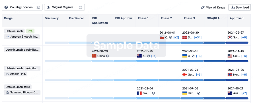
Biosimilar
Competitive landscape of biosimilars in different countries/locations. Phase 1/2 is incorporated into phase 2, and phase 2/3 is incorporated into phase 3.
login
or
Regulation
Understand key drug designations in just a few clicks with Synapse.
login
or

AI Agents Built for Biopharma Breakthroughs
Accelerate discovery. Empower decisions. Transform outcomes.
Get started for free today!
Accelerate Strategic R&D decision making with Synapse, PatSnap’s AI-powered Connected Innovation Intelligence Platform Built for Life Sciences Professionals.
Start your data trial now!
Synapse data is also accessible to external entities via APIs or data packages. Empower better decisions with the latest in pharmaceutical intelligence.
Bio
Bio Sequences Search & Analysis
Sign up for free
Chemical
Chemical Structures Search & Analysis
Sign up for free