Request Demo
Last update 09 Dec 2025

Université Paris Diderot-Paris 7
Last update 09 Dec 2025
Overview
Basic Info
Introduction Located in the heart of Paris, France, Paris Diderot University, previously known as Paris 7, is a prestigious institution of higher education. As one of the successors to the historic University of Paris, which was disbanded in 1970, Paris Diderot University was established by a group of professors from various faculties, including Science, Medicine, and Humanities. The university was renamed after the renowned French philosopher, art critic, and writer Denis Diderot in 1994. |
Corporation Tree
Boost your research with our corporation tree data.
login
or

Pipeline
Pipeline Snapshot as of 19 Dec 2025
The statistics for drugs in the Pipeline is the current organization and its subsidiaries are counted as organizations,Early Phase 1 is incorporated into Phase 1, Phase 1/2 is incorporated into phase 2, and phase 2/3 is incorporated into phase 3
Other
3
Login to view more data
Current Projects
Login to view more data
Deal
Boost your decision using our deal data.
login
or

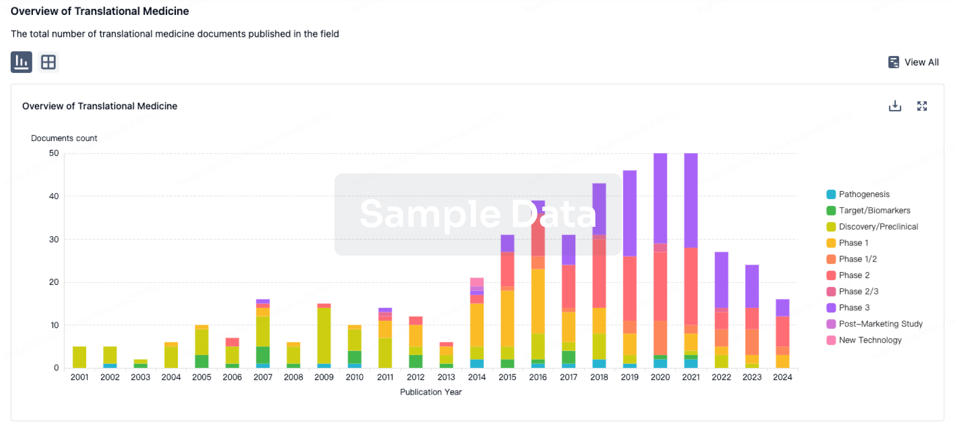
Translational Medicine
Boost your research with our translational medicine data.
login
or

Profit
Explore the financial positions of over 360K organizations with Synapse.
login
or

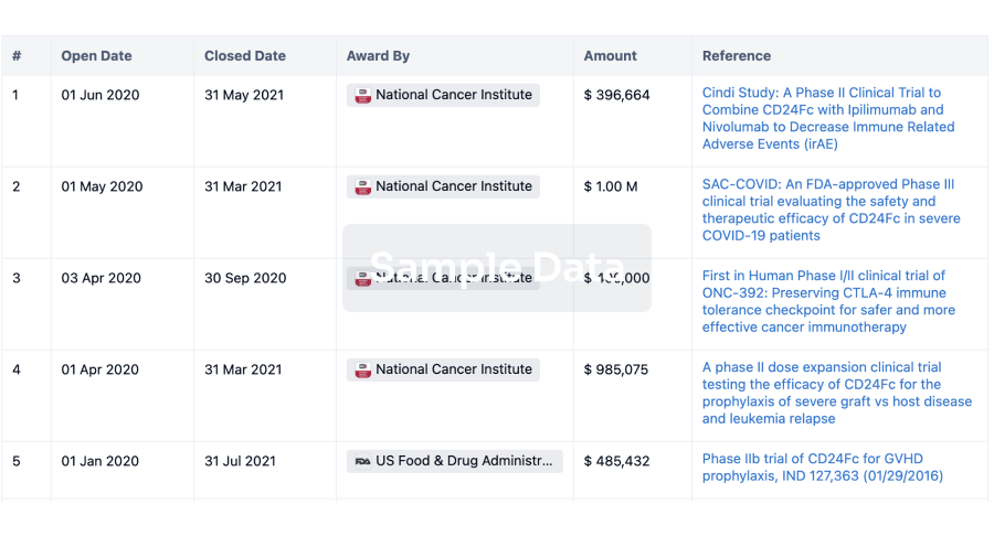
Grant & Funding(NIH)
Access more than 2 million grant and funding information to elevate your research journey.
login
or

Investment
Gain insights on the latest company investments from start-ups to established corporations.
login
or

Financing
Unearth financing trends to validate and advance investment opportunities.
login
or

AI Agents Built for Biopharma Breakthroughs
Accelerate discovery. Empower decisions. Transform outcomes.
Get started for free today!
Accelerate Strategic R&D decision making with Synapse, PatSnap’s AI-powered Connected Innovation Intelligence Platform Built for Life Sciences Professionals.
Start your data trial now!
Synapse data is also accessible to external entities via APIs or data packages. Empower better decisions with the latest in pharmaceutical intelligence.
Bio
Bio Sequences Search & Analysis
Sign up for free
Chemical
Chemical Structures Search & Analysis
Sign up for free