Request Demo
What is the mechanism of FLUORODOPA F-18?
17 July 2024
Fluorodopa F-18, also known as 18F-DOPA, is a radiopharmaceutical commonly utilized in positron emission tomography (PET) imaging to assess various neurological conditions, particularly Parkinson's disease and other movement disorders. Its mechanism of action is deeply rooted in its biochemical similarities to the naturally occurring amino acid, L-DOPA, which is a critical precursor in the biosynthesis of the neurotransmitter dopamine. Understanding the mechanism of Fluorodopa F-18 requires a comprehensive look at its pharmacokinetics and interaction with the dopaminergic system.
The journey of Fluorodopa F-18 begins with its administration, typically via intravenous injection. Upon entering the bloodstream, it is transported across the blood-brain barrier through specific amino acid transporters located on the endothelial cells lining the cerebral vasculature. This selective transport is facilitated by the structural resemblance of 18F-DOPA to L-DOPA, allowing it to exploit the same biological pathways.
Once inside the brain, 18F-DOPA undergoes a series of biochemical transformations much like its natural counterpart. The first critical step involves its conversion to fluorodopamine by the enzyme aromatic L-amino acid decarboxylase (AADC), also known as DOPA decarboxylase. This conversion predominantly occurs in the presynaptic neurons of the striatum, a key region involved in motor control.
Fluorodopamine, the active metabolite of 18F-DOPA, is then sequestered into synaptic vesicles via the vesicular monoamine transporter (VMAT2). This storage mimics the normal physiological storage of dopamine, positioning it for eventual release into the synaptic cleft. Upon neuronal activation, fluorodopamine is released into the synaptic cleft where it can bind to dopamine receptors on postsynaptic neurons, thereby participating in dopaminergic neurotransmission.
The PET imaging capabilities of Fluorodopa F-18 arise from its radioactive fluorine-18 isotope, which emits positrons upon decay. These positrons quickly annihilate with electrons in the surrounding tissue, producing pairs of gamma photons that travel in nearly opposite directions. PET scanners detect these gamma photons, enabling the construction of precise images that reflect the distribution and concentration of 18F-DOPA within the brain.
The distribution pattern of Fluorodopa F-18 is particularly informative in diagnosing and understanding Parkinson's disease. In patients with Parkinson's, there is a marked reduction in dopaminergic neurons within the substantia nigra and their terminals in the striatum. PET imaging with 18F-DOPA reveals this deficit through decreased uptake and conversion of 18F-DOPA in the striatum, providing valuable diagnostic and prognostic information.
Moreover, Fluorodopa F-18 PET imaging is not limited to Parkinson's disease. It is also utilized in the evaluation of other movement disorders, certain types of brain tumors such as pheochromocytomas and paragangliomas, and even in some psychiatric disorders where dopaminergic dysfunction is implicated.
In summary, the mechanism of Fluorodopa F-18 hinges on its biochemical similarity to L-DOPA, enabling its transport into the brain, conversion to fluorodopamine, and participation in the dopaminergic system. Its diagnostic utility is primarily derived from the radioactive decay of fluorine-18, which facilitates detailed PET imaging of dopaminergic function. This makes 18F-DOPA an invaluable tool in the diagnosis and management of neurological conditions involving dopaminergic dysfunction.
How to obtain the latest development progress of all drugs?
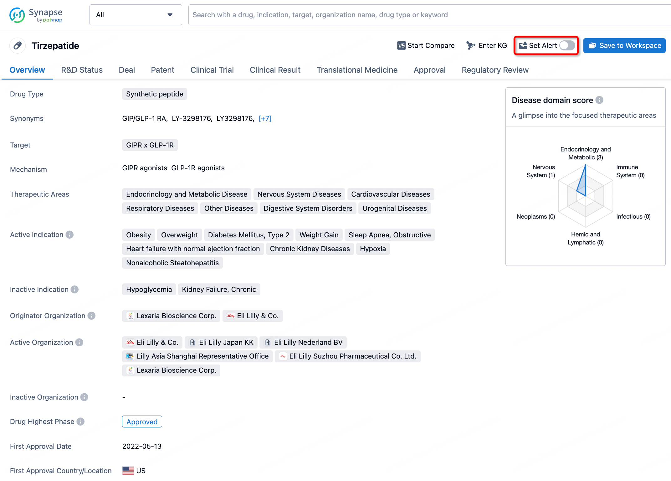
In the Synapse database, you can stay updated on the latest research and development advances of all drugs. This service is accessible anytime and anywhere, with updates available daily or weekly. Use the "Set Alert" function to stay informed. Click on the image below to embark on a brand new journey of drug discovery!


AI Agents Built for Biopharma Breakthroughs
Accelerate discovery. Empower decisions. Transform outcomes.
Get started for free today!
Accelerate Strategic R&D decision making with Synapse, PatSnap’s AI-powered Connected Innovation Intelligence Platform Built for Life Sciences Professionals.
Start your data trial now!
Synapse data is also accessible to external entities via APIs or data packages. Empower better decisions with the latest in pharmaceutical intelligence.