Rodent Optic Nerve Head Wins the 49th Annual Nikon Small World Photo Microscopy Competition
17 Oct 2023
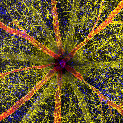
The 2023 first place image showcases a debilitating and prevalent complication of diabetes
MELVILLE, N.Y., Oct. 17, 2023 /PRNewswire/ -- Nikon Instruments Inc. today unveiled the winners of the 49th annual Nikon Small World Photomicrography Competition. This year's first place prize was awarded to Hassanain Qambari, assisted by Jayden Dickson of the Lions Eye Institute, for his vivid image of a rodent optic nerve head showing astrocytes (yellow), contractile proteins (red), and retinal vasculature (green). The colorful image provides an important contribution to the study and reversal of diabetic retinopathy, which affects one in five persons with diabetes worldwide.1
Continue Reading

Preview
Source: PRNewswire
Nikon Small World (PRNewsfoto/Nikon Instruments Inc.)
Preview
Source: PRNewswire
Rodent optic nerve head showing astrocytes (yellow), contractile proteins (red) and retinal vasculature (green)
Diabetic retinopathy occurs when high blood sugar damages the blood vessels in the tissue at the back of the eye, known as the retina. The damaged blood vessels can swell and leak, which can cause blurry vision or total loss of eyesight. Since 2021, Qambari has devoted his time and research to the early detection and reversal of the disease.
"Current diagnostic criteria and treatment regimens for diabetic retinopathy are limited to the late-stage appearance of the disease, with irreversible damage to retinal microvasculature and function," said Qambari. "The visual system is a complex and highly specialized organ, with even relatively minor perturbations to the retinal circulation able to cause devastating vision loss. I entered the competition as a way to showcase the complexity of retinal microcirculation."
Qambari faced some challenges when capturing his image such as locating fine vessels near 110 microns in diameter and establishing a protocol for labeling different cell types. "Over the past 20 years, our research group has refined the technique of isolated ocular perfusion labeling for fine vessels in the eye," said Qambari. "While the ophthalmic artery in the rodent model presented a technically demanding challenge, we were able to overcome it with persistence and patience."
The final result educates the public about the universal condition affecting millions and the vital research necessary to further advance care. "The Nikon Small World competition is great, as it showcases amazing work across many disciplines from around the world," said Qambari. "All the images presented in the competition represent the beauty and artistic side of science which may otherwise get overlooked. Such a competition not only celebrates the participant's hard work and passion but may also draw and inspire young scientists to pursue a career in STEM. It certainly inspired me."
Like Qambari, Eric Flem, Senior Manager, CRM and Communications at Nikon Instruments, is passionate about sharing exemplary scientific work and artistic techniques. "The past 49 years of this competition have borne witness to many innovative and pioneering advancements in scientific imaging technology," said Flem. "I am consistently awed by how these advancements make it possible to create art out of science for the public to enjoy."
Second place was awarded to Ole Bielfeldt for his image of a matchstick igniting by the friction surface of the box. The image was taken within one eight-thousands of a second and utilized imaging stacking.
Third place was awarded to Malgorzata Lisowska for her image of breast cancer cells.
In addition to the top three winners, Nikon Small World recognized 83 photos out of thousands of entries from scientists and artists across the globe.
The 2023 judging panel included:
Ed Cara, Science and Health Reporter at Gizmodo
James Cutmore, Picture Editor at BBC Science Focus Magazine
Dr. Gary Laevsky, Director of the Confocal Imaging Facility at Princeton University
Dr. Igor Siwanowicz, Research Scientist at Howard Hughes Medical Institute
Dr. Clare Waterman, Cell Biologist and Member of the National Academy of Sciences
For additional information, please visit www.nikonsmallworld.com, or follow the conversation on Facebook, Twitter @NikonSmallWorld and Instagram @NikonInstruments.
NIKON SMALL WORLD WINNERS
1st Place
Hassanain Qambari & Jayden Dickson
Department of Physiology & Pharmacology
Perth, Western Australia, Australia
Rodent optic nerve head showing astrocytes (yellow), contractile proteins (red) and retinal vasculature (green)
Confocal, Fluorescence, Image Stacking
20X (Objective Lens Magnification)
2nd Place
Ole Bielfeldt
Macrofying
Cologne, North Rhine-Westphalia, Germany
Matchstick igniting by the friction surface of the box.
Brightfield, Image Stacking
2.5X (Objective Lens Magnification)
3rd Place
Malgorzata Lisowska
Independent Value Based Healthcare Consultant
Warsaw, Mazowieckie, Poland
Breast cancer cells
Brightfield, Image Stacking
40X (Objective Lens Magnification)
4th Place
John-Oliver Dum
Medienbunker Produktion
Bendorf, Rheinland-Pfalz, Germany
Venomous fangs of a small tarantula
Image Stacking
10X (Objective Lens Magnification)
5th Place
Dr. David Maitland
www.davidmaitland.com
Feltwell, Norfolk, United Kingdom
Auto-fluorescing defensive hairs covering the leaf surface of Eleagnus angustifolia exposed to UV light
Fluorescence, Image Stacking
10X (Objective Lens Magnification)
6th Place
Timothy Boomer
WildMacro.com
Vacaville, California, USA
Slime mold (Comatricha nigra) showing capillitial fibers through its translucent peridium
Image Stacking
10X (Objective Lens Magnification)
7th Place
Dr. Grigorii Timin & Dr. Michel Milinkovitch
Department of Genetics and Evolution
Geneva, Switzerland
Mouse embryo
Light Sheet
4X (Objective Lens Magnification)
8th Place
Stefan Eberhard
Athens, Georgia, USA
Caffeine crystals
Polarized Light
25X (Objective Lens Magnification)
9th Place
Vaibhav Deshmukh
Department of Molecular Physiology and Biophysics
Houston, Texas, USA
Cytoskeleton of a dividing myoblast; tubulin (cyan), F-actin (orange) and nucleus (magenta)
Fluorescence, Structured Illumination Microscopy (SIM)
63X (Objective Lens Magnification)
10th Place
Melinda Beccari & Dr. Don W. Cleveland
Department of Cellular and Molecular Medicine
La Jolla, California, USA
Motor neurons grown in microfluidic device for separation of cell bodies (top) and axons (bottom). Green - microtubules; Red - growth cones (actin).
Confocal, Fluorescence
20X (Objective Lens Magnification)
11th Place
Dr. Diego García
Real Sociedad Española de Física
Madrid, Spain
Crystallized sugar syrup
Polarized Light
25X (Objective Lens Magnification)
12th Place
Sherif Abdallah Ahmed
Department of Zoology
Tanta, Egypt, Arab Republic
"Cuckoo wasp" standing on a flower
Image Stacking
4X (Objective Lens Magnification)
13th Place
Satu Paavonsalo & Dr. Sinem Karaman
Individualized Drug Therapy Research Program, Faculty of Medicine
Helsinki, Finland
Blood and lymphatic vasculatures in the ear skin of an adult mouse
Confocal
10X (Objective Lens Magnification)
14th Place
John-Oliver Dum
Medienbunker Produktion
Bendorf, Rheinland-Pfalz, Germany
Sunflower pollen on an acupuncture needle
Image Stacking
40X (Objective Lens Magnification)
15th Place
Dr. Pichaya Lertvilai
Scripps Institution of Oceanography
La Jolla, California, USA
Fluorescent image of an Acropora sp. showing individual polyps with symbiotic zooxanthellae
Darkfield, Fluorescence, Image Stacking
5X (Objective Lens Magnification)
16th Place
Dr. Diego García
Real Sociedad Española de Física
Madrid, Spain
Carbon nanotubes
Stereomicroscopy
30X (Objective Lens Magnification)
17th Place
Yuan Ji
World Expo Museum
Shanghai, China
Chinese moon moth (Actias ningpoana) wing scales
Image Stacking
20X (Objective Lens Magnification)
18th Place
Scott Peterson
New Hope, Minnesota, USA
A cryptocrystalline micrometeorite resting on a #80 testing sieve.
Image Stacking
20X (Objective Lens Magnification)
19th Place
Marek Miś
Marek Miś Photography
Suwalki, Podlaskie, Poland
Stomata in peace lily (Spathiphyllum sp.) leaf epidermis
Polarized Light
40X (Objective Lens Magnification)
20th Place
Daniel Castranova & Dr. Brant Weinstein
Bethesda, Maryland, USA
Adult transgenic zebrafish head showing blood vessels (blue), lymphatic vessels (yellow), and the skin and scales (magenta)
Confocal
4X (Objective Lens Magnification)
HM
Dr. Arthur Chien & Dr. Ann Na Cho
Microscopy Facility
Macquarie University, New South Wales, Australia
Organ-on-chip system enabling the synaptic conjugation between 3D human embryonic stem cells
Confocal
10X (Objective Lens Magnification)
HM
Dr. Amy Engevik
Department of Regenerative Medicine & Cell Biology
Charleston, South Carolina, USA
Neonatal mouse intestinal tissue cells
Fluorescence
20X (Objective Lens Magnification)
HM
Dr. Nathan P. Myhrvold
Modernist Cuisine
Bellevue, Washington, USA
Trichinella cyst in pork muscle (Trichinella is a parasitic worm known to cause trichinosis)
Brightfield, Image Stacking
10X (Objective Lens Magnification)
HM
Ángel Navarro Gómez
Madrid, Spain
Carpenter bee (Xylocopa violacea) head and antenna
Image Stacking
20X (Objective Lens Magnification)
HM
Dr. Andrew M. Posselt
Division of Transplant Surgery
Mill Valley, California, USA
Underside of cellar spider (Pholcus phalangioides)
Image Stacking
10X (Objective Lens Magnification)
HM
Dr. Grigorii Timin & Dr. Michel Milinkovitch
Department of Genetics and Evolution
Geneva, Switzerland
Dermal collagen in embryonic snake scales
Confocal
63X (Objective Lens Magnification)
HM
Dr. Bas van Bommel
Department of Biochemistry
Berlin, Germany
Rat astrocytes
Confocal
40X (Objective Lens Magnification)
HM
Travis Wagner
Department of Mechanical Engineering
Rochester, New York, USA
Sphagnum moss with two air bubbles on the sample
Fluorescence, Image Stacking
20X (Objective Lens Magnification)
IoD
Dr. Florian Alonso
BioTis-INSERM U1026
Pessac, Gironde, France
Formation of blood vessels (angiogenesis) in the retina from a Lifeact-EGFP newborn mouse
Confocal
63X (Objective Lens Magnification)
IoD
Raghuram Annadana
Raghuram Annadana Photography
Bangalore, Karnataka, India
Developing stamen and stigma inside a Hibiscus flower bud
Panoramic Image Stacking
10X (Objective Lens Magnification)
IoD
Dr. Alexandre Beber
Vestec, Central Bohemia, Czech Republic
Fluorescent actin filaments (yellow) and fluorescent anillin protein (blue) deposited on a glass coverslip after dewetting
Fluorescence, Confocal
60X (Objective Lens Magnification)
IoD
Todd Becker
Teabeck
Verona, Wisconsin, USA
Platinum spark plug
Image Stacking, Reflected Light
4X (Objective Lens Magnification)
IoD
Dr. Frantisek Bednar
Svosov, Zilinsky, Slovak Republic
Slime mold (Trichia crateriformis)
Image Stacking
5X (Objective Lens Magnification)
IoD
Taylor Bell
Gustometry + SF Micro Society
Norwalk, Connecticut, USA
Freshwater amphipod
Image Stacking
4X (Objective Lens Magnification)
IoD
Timothy Boomer
WildMacro.com
Vacaville, California, USA
Slime mold (Didymium sp.) fruiting bodies
Image Stacking
10X (Objective Lens Magnification)
IoD
Dr. Michael John Bridge & Michael Sieverts
HSC Cell Imaging Core
Salt Lake City, Utah, USA
Mouse femur bone lacunar-canalicular network (voids in bone that house osteocytes and their interconnected micro-tubular processes)
Confocal
63X (Objective Lens Magnification)
IoD
Arturo Calderón, Dr. Miguel Tapia-Rodríguez & Dr. Juan Pedro Laclette
Department of Immunology
Mexico City, Mexico
Muscle architecture of an evaginating tapeworm (Taenia crassiceps cysticercus)
Confocal, Fluorescence, Image Stacking
10X (Objective Lens Magnification)
IoD
Hsuan Chen
Chen-Hui Chen's Lab in the Institute of Cellular and Organismic Biology
Taipei City, Taipei, Taiwan
In toto image of the skin and mucous cells in a live zebrafish larva
Confocal
10X (Objective Lens Magnification)
IoD
Dr. Arthur Chien
Microscopy Facility
Macquarie University, New South Wales, Australia
Cleared mouse embryo
Stereomicroscopy, Reflected Light, Focus Stacking
0.5X (Objective Lens Magnification)
IoD
Nikky Corthout & Alex Calzoni
Leuven, Vlaams-Brabant, Belgium
Retrograde labeled neurons in the cortex of a cortical mouse brain section
Image Stacking, Fluorescence, Confocal
20X (Objective Lens Magnification)
IoD
John-Oliver Dum
Medienbunker Produktion
Bendorf, Rheinland-Pfalz, Germany
Cabbage butterfly eggs
Brightfield, Image Stacking
20X (Objective Lens Magnification)
IoD
Nadia Efimova
Philadelphia, Pennsylvania, USA
Maturing mouse cortical neuron in culture
Confocal, Deconvolution, Fluorescence
63X (Objective Lens Magnification)
IoD
Ricardo Roberto Fernández Martínez
IES Virgen de la Luz
Department of Biology and Geology
Avilés, Asturias, Spain
Tail of planktonic shrimp larvae
Image Stacking
17X (Objective Lens Magnification)
IoD
Frank Fox
Konz, Rheinland-Pfalz, Germany
Marine organism (Pyrocystis lunula, Dinophyceae)
Differential Interference Contrast (DIC)
100X (Objective Lens Magnification)
IoD
Ian Gardiner
Calgary, Alberta, Canada
Clam shrimp (Lynceus mucronatus)
Reflected Light, Focus Stacking
2.5X (Objective Lens Magnification)
IoD
Dr. Saikat Ghosh & Dr. Juan S. Bonifacino
NICHD
Bethesda, Maryland, USA
iPSC-derived human neurons
Confocal
40X (Objective Lens Magnification)
IoD
Dr. Thomas G.W. Graham
Department of Molecular and Cell Biology
Berkeley, California, USA
Algae from a mud puddle
Confocal
10X (Objective Lens Magnification)
IoD
Daniel B. Hoffman & Dr. Jacob Sorensen
Department of Kinesiology
Minneapolis, Minnesota, USA
Rat skeletal muscle fibers with associated neuromuscular junctions (white)
Confocal
40X (Objective Lens Magnification)
IoD
Dr. Hema Saranya Ilamathi
Department of Medical biology
Trois-Rivieres, Quebec, Canada
Distribution of cellular batteries (mitochondria-yellow), along the transport cables (Tubulin-red, actin-cyan) in human fibroblast
Confocal
63X (Objective Lens Magnification)
IoD
Don Komarechka
Komarechka Photography Ltd.
Ravna Gora, Varna, Bulgaria
Two fluorescing diamonds
Fluorescence
10X (Objective Lens Magnification)
IoD
Charles Krebs
Charles Krebs Photography
Issaquah, Washington, USA
Feeding bryozoan colony zooids. Bryozoans are microscopic aquatic invertebrates that live in colonies.
Reflected Light
5X (Objective Lens Magnification)
IoD
Charles Krebs
Charles Krebs Photography
Issaquah, Washington, USA
Mushroom gills showing sporophores (sporangiophores)
Darkfield, Image Stacking
20X (Objective Lens Magnification)
IoD
Dr. Håkan Kvarnström
Bromma, Sweden
Amoeba (Arcella)
Differential Interference Contrast (DIC)
60X (Objective Lens Magnification)
IoD
Michael Landgrebe
Weissensberg, Bavaria, Germany
Fossil diatom
Brightfield (inverted)
60X (Objective Lens Magnification)
IoD
Dr. Francisco Lázaro-Diéguez
Bronx, New York, USA
Non-parenchymal liver cells
Confocal
63X (Objective Lens Magnification)
IoD
Dr. Olivier Leroux
Gent, Oost-Vlaanderen, Belgium
Composition of transverse sections of plant organs
Fluorescence
20X (Objective Lens Magnification)
IoD
Dr. Pichaya Lertvilai
Scripps Institution of Oceanography
La Jolla, California, USA
Coral (Acropora granulosa) fluorescing under blue light
Image Stacking, Fluorescence, Darkfield
5X (Objective Lens Magnification)
IoD
Dr. Yu-Hsiu Liu
Chen-Hui Chen's Lab in the Institute of Cellular and Organismic Biology
Nankang, Taipei, Taiwan
Palmskin zebrafish larva
Confocal
25X (Objective Lens Magnification)
IoD
Walter Machielsen
www.waltermachielsen.com
Rotterdam, Zuid-Holland, Netherlands
Buckthorn trichomes
Differential Interference Contrast (DIC)
40X (Objective Lens Magnification)
IoD
Dr. David Maitland
www.davidmaitland.com
Feltwell, Norfolk, United Kingdom
Wing scales of the cinnabar moth (Tyria jacobaeae) under ultraviolet light (UV)
Fluorescence, Image Stacking
20X (Objective Lens Magnification)
IoD
Sébastien Malo
Saint Lys, Haute-Garonne, France
Crab spider (Thomisus onustus)
Darkfield, Image Stacking, Reflected Light
6.3X (Objective Lens Magnification)
IoD
Sébastien Malo
Saint Lys, Haute-Garonne, France
Geranium (Geraniaceae) stamen covered in pollen
Darkfield, Image Stacking, Reflected Light
6.3X (Objective Lens Magnification)
IoD
Amir Maqbool
Higher Education Department Jammu and Kashmir India
Department of Zoology
Srinagar, Jammu and Kashmir, India
Phoretic mites on the leg of a bumblebee
Image Stacking
3X (Objective Lens Magnification)
IoD
Dr. Robert Markus
School of Life Sciences Imaging (SLIM)
Nottingham, Nottinghamshire, United Kingdom
Actin cytoskeleton of bovine pulmonary epithelial cells
Structured Illumination Microscopy (SIM)
63X (Objective Lens Magnification)
IoD
Dr. Simon Frederik Merz, Dr. Lea Bornemann & Dr. Ewa Patrycja Smajek
Department of Biophysics
Bielefeld, North-Rhine Westphalia, Germany
Fly (cyan) caught in a Venus flytrap (red)
Fluorescence, Light Sheet
2.4X (Objective Lens Magnification)
IoD
Marek Miś
Marek Miś Photography
Suwalki, Podlaskie, Poland
Leaf epidermis stomata (Stromanthe sp.)
Polarized Light
10X (Objective Lens Magnification)
IoD
Ángel Navarro Gómez
Madrid, Spain
Mechanosensors in a Venus flytrap
Image Stacking
20X (Objective Lens Magnification)
IoD
Dr. Foo Yong Ng
Faculty of Science and Technology, Universiti Kebangsaan Malaysia
Department of Biology Science and Biotechnology
Bangi, Selangot, Malaysia
Moss
Image Stacking
2X (Objective Lens Magnification)
IoD
Dr. Lori O'Brien
Department of Cell Biology and Physiology
Chapel Hill, North Carolina, USA
Embryonic mouse (Mus musculus) kidney showing the collecting duct (blue) and nephron progenitor (yellow) cells
Confocal
5X (Objective Lens Magnification)
IoD
Yusuf Ziya Öztürk
TCDD Teknik Müh. Müş. A.Ş.
Ankara, Çankaya, Turkey
Bee
Image Stacking
3.7X (Objective Lens Magnification)
IoD
Alison Pollack
San Anselmo, California, USA
Slime mold (Craterium leucocephalum), looking like a beautiful tiny goblet
Image Stacking, Reflected Light
10X (Objective Lens Magnification)
IoD
Alison Pollack
San Anselmo, California, USA
Slime mold (Diderma tigrinum)
Image Stacking, Reflected Light
10X (Objective Lens Magnification)
IoD
Dr. Andrew M. Posselt
Division of Transplant Surgery
Mill Valley, California, USA
Blue black weevil (Metapocyrtus sp.)
Image Stacking
6X (Objective Lens Magnification)
IoD
Frank Reiser
Department of Biology
Garden City, New York, USA
Ostracods and algae (Cladophora)
Darkfield, Reflected Light
2.5X (Objective Lens Magnification)
IoD
Dr. Colin Rogers & Dr. Erica Weekman
Sanders Brown Center on Aging
Lexington, Kentucky, USA
Mouse retina blood vessels (green) astrocytes (red) and microglia (purple)
Confocal
20X (Objective Lens Magnification)
IoD
Jan Rosenboom
Rostock, Mecklenburg Vorpommern, Germany
Diatoms (single-celled algae) arranged on the head of a pin
Image Stacking, Reflected Light
4X (Objective Lens Magnification)
IoD
Danny Sanchez
Mineralien LLC
Valley Village, California, USA
Golden rutile in quartz
Reflected Light, Darkfield
6X (Objective Lens Magnification)
IoD
Hans Schoofs
Department of Immunology, Genetics and Pathology (IGP)
Uppsala, Sweden
Blood and lymphatic vessels in a mouse diaphragm
Confocal
20X (Objective Lens Magnification)
IoD
Dr. Leo Serra
Cambridge, Cambridgeshire, United Kingdom
Bindweed (Convolvulus) leaf epidermal cells autofluorescing
Confocal
20X (Objective Lens Magnification)
IoD
Dr. Leo Serra
Cambridge, Cambridgeshire, United Kingdom
Patterns at the surface of an embryonic leaf of Thale cress (Arabidopsis thaliana)
Confocal
20X (Objective Lens Magnification)
IoD
Stephen Vidman & Dr. Andrea Tedeschi
Department of Neuroscience
Columbus, Ohio, USA
3D capillary network section of the mammalian brain (dentate gyrus of the hippocampus)
Confocal, Deconvolution
10X (Objective Lens Magnification)
IoD
Priscilla Vieto Bonilla & Brandon Antonio Segura Torres
Department of Biological Sciences
San Carlos de Bariloche, Río Negro, Argentina
One-week-old Axolotl after hatching
Image Stacking, Stereomicroscopy
25X (Objective Lens Magnification)
IoD
Cagri Yalcin
Impressions Microscopiques
Amsterdam, Noord Holland, Netherlands
Crystals of malonic acid dissolved in ethanol
Polarized Light
4X (Objective Lens Magnification)
IoD
Zhao Zengchao
Hefei, Anhui Province, China
Bristle of a millipede (Polyxenidae)
Image Stacking
50X (Objective Lens Magnification)
IoD
Dr. Tong Zhang
Biological Imaging Facility
Evanston, Illinois, USA
Lily (Lilium) anther cross section with pollen
Confocal
20X (Objective Lens Magnification)
IoD
Dr. Tong Zhang
Biological Imaging Facility
Evanston, Illinois, USA
Lily (Lilium) anther cross section with pollen
Confocal
20X (Objective Lens Magnification)
About Nikon Small World Photomicrography Competition
The Nikon Small World Competition is open to anyone with an interest in photography or video. In 2024, the competition will celebrate its 50th anniversary. Ahead of the celebration, participants may upload digital images and videos directly at www.nikonsmallworld.com. For additional information, contact Nikon Small World, Nikon Instruments Inc., 1300 Walt Whitman Road, Melville, NY 11747, USA, or phone (631) 547-8569. Entry forms for Nikon's 2024 Small World and Small World in Motion Competitions are available at https://enter.nikonsmallworld.com/
About Nikon Instruments Inc.
Nikon Instruments Inc. is the US microscopy arm of Nikon Healthcare, a world leader in the development and manufacture of optical and digital imaging technology for biomedical applications. For more information, visit https://www.microscope.healthcare.nikon.com/ or contact us at 1-800-52-NIKON.
1 Global Prevalence of Diabetic Retinopathy and Projection of Burden through 2045: Systematic Review and Meta-analysis
Teo Z.L., Tham Y.-C., Yu M., Chee M.L., Rim T.H., Cheung N., Bikbov M.M., (...), Cheng C.-Y.
(2021) Ophthalmology, 128 (11) , pp. 1580-1591.
SOURCE Nikon Instruments Inc.
For more details,please visit the original website
The content of the article does not represent any opinions of Synapse and its affiliated companies. If there is any copyright infringement or error, please contact us, and we will deal with it within 24 hours.
Organizations
Indications
Drugs
-Hot reports


Get started for free today!
Accelerate Strategic R&D decision making with Synapse, PatSnap’s AI-powered Connected Innovation Intelligence Platform Built for Life Sciences Professionals.
Start your data trial now!
Synapse data is also accessible to external entities via APIs or data packages. Leverages most recent intelligence information, enabling fullest potential.